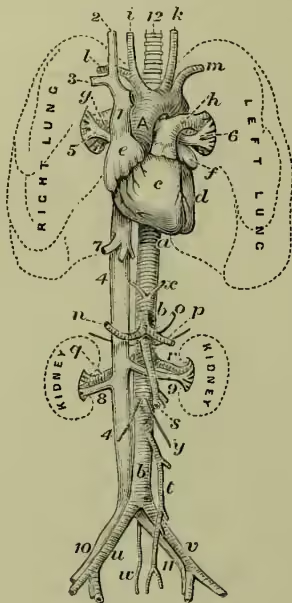
Aorta is the great arterial trunk which, rising from the left ventricle of the heart, sends its branches ramifying through the whole body. The aorta in man is subdivided by anatomists into the arch, the thoracic aorta, and the abdominal aorta. The arch is a loop with the convexity directed upwards, forwards, and to the right side, reaching at its highest part to a level with the upper border of the cartilage of the second rib. Changing its direction, it runs from right to left, and from before backwards to reach the left side of the spine, with which it first comes in contact between the fourth and fifth dorsal vertebrae. Here it descends, and at the lower border of the fifth dorsal vertebra it becomes the thoracic aorta. Five arteries arise from the arch—viz. two coronaries, for the supply of the muscular tissue of the heart itself; the innominate, dividing into the right common carotid and right subclavian arteries; and the left common carotid and left subclavian arteries. 1, ascending part of the arch of the aorta; a, thoracic aorta; bb, abdominal aorta; c, d, right and left ventricles of heart; e, f, right and left auricles of the heart; g, h, right and left pulmonary arteries; i, k, right and left common carotid arteries; l, m, right and left subclavian arteries; n, hepatic artery; o, gastric artery; p, splenic artery; q, r, right and left renal arteries; s, t, superior and inferior mesenteric arteries; u, v, right and left common iliac arteries; w, middle sacral artery; x, phrenic arteries; y, spermatic arteries.
2, superior vena cava; 3, right internal jugular vein; 4, right subclavian vein (the left is removed to show the arch of the aorta); 5, inferior vena cava; 6, 8, right and left pulmonary veins; 7, hepatic veins; 9, 11, right and left renal veins; 10, 11, right and left iliac veins; 12, trachea.
At the commencement of the arch are three small swellings or pouches, the aortic sinuses, internal to which are the three semilunar valves or folds of the lining membrane, which prevent regurgitation of the blood into the heart. The thoracic aorta extends from the lower border of the fifth dorsal vertebra to the diaphragm, gradually getting into the middle line of the spine. The thoracic aorta gives off the bronchial arteries (two or three) to supply the tissue of the lungs; and some small branches (three or four) to the oesophagus, and intercostal arteries, to supply the walls of the chest (usually nine on each side, the upper two intercostal spaces being supplied by a branch from the subclavian artery). The abdominal aorta passes from the diaphragm to the fourth lumbar vertebra, opposite the body of which it divides into the two common iliac trunks. The abdominal aorta gives off: (a) Branches to the abdominal viscera. These are either single or in pairs. The single arteries are—the coeliac axis, which divides into three large branches for the stomach, liver, and spleen; the superior mesenteric, for the small, and part of the large intestine; the inferior mesenteric, for the large intestine not supplied by the superior mesenteric. The paired arteries are the supra-renal, renal, and spermatic or ovarian. (b) Branches to the abdominal walls—viz. the two phrenic arteries to the diaphragm; the lumbar arteries, four or five pairs, for the supply of the lower part of the abdominal wall (the loins); the middle sacral or caudal artery, a small vessel which arises from the back of the aorta just above its point of bifurcation. In fish and animals with tails, this branch is the caudal prolongation of the aorta.
The above is the usual arrangement; but occasionally it varies, especially in the number of arteries springing from the arch. The structure of the aorta will be given under ARTERIES; and the comparative anatomy under CIRCULATION.
During foetal life, there is a communication between the arch of the aorta and the pulmonary artery, called the ductus arteriosus, the canal of which becomes obliterated after birth. In the human carotid artery, it has been calculated that the blood-stream moves with a velocity of inches per second, and that the pressure of blood in the same vessel would support a weight of 3 lb. 2 oz. to the square inch.
The coats of the aorta are very subject to fatty disease termed Atheroma (q.v.), and in advanced life, to calcareous degeneration or deposit of lime-salts, which destroys their elasticity. This change renders them very liable to Aneurysm (q.v.), which, as may be expected, is generally situated at the curves of the aorta, especially at the arch, where the force of the blood-stream is greatest.