Ascidians, or TUNICATES, are a class of degenerate survivors of ancestral vertebrates. They were observed and well characterised by Aristotle, but were never really understood till 1866, when the discovery of their complete life-history warranted naturalists in removing them from their random position beside molluscs to the base of the vertebrate series. The larvæ exhibit unquestionable vertebrate characters—viz. a spinal cord, brain, and cerebral eye, a notochord (q.v.), gill-slits, ventral heart, &c. Except in Appendicularia, however, the vertebrate characters of the larvæ are to a large extent lost in the usually sessile degenerate adult. The tail shrivels up, the notochord and spinal cord disappear, the brain is represented merely by a small ganglion, and the whole symmetry of the body is altered. The ordinary ascidians, the sea-squirts, so called from their power of jerking out water when touched, are common along the coasts of all seas, ranging from low water to twenty fathoms, and usually attached to stones or shells, while some of the more complex forms are free-swimming. Few of them measure separately more than 3 or 4 inches, and most of them decidedly less, but the more or less intimate colonies frequently formed often attain larger dimensions. Chains of free-swimming ascidians sometimes occur several feet in length.
The following account refers primarily to a simple ascidian, and ought to be prefaced by reading the account of Appendicularia (q.v.), which, as has been noted, retains the larval characters lost in other ascidians. The animal has a double-mouthed flask shape, and is enveloped in a transparent, gelatinous, external sheath, which is secreted by the real skin. This test is remarkable as containing Cellulose (q.v.), the substance which forms the cell-wall of vegetable organisms. Through one of the apertures which are usually fringed with minute lobes, and often bear little coloured spots, the water enters the body; through the other the water, faeces, and reproductive elements are expelled. The anterior region of the alimentary caual forms a large respiratory pharynx, such as also occurs in one worm-like form, Balanoglossus, in Amphioxus (q.v.), and in all vertebrates in the embryonic stage at least. As in Appendicularia, a pair of involutions from the skin meet outgrowths from the gut, and establish a primitive respiratory communication with the exterior. In
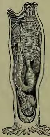
Structure of a simple Ascidian, showing inhalent aperture, leading into respiratory pharynx; looped alimentary canal, opening along with genital duct into cloacal chamber; nerve ganglion between inhalent and exhalent apertures; reproductive organs near the base, eggs in body-cavity, &c.; heart at very base; fixing processes. (After Hæckel.) the ascidian, however, this simple structure is replaced, as afterwards described, by a multitude of gill-slits opening from the pharynx into a surrounding chamber which communicates with the exterior by a single aperture. The water is drawn in by the mouth, passes through the richly ciliated slits of the pharynx, bathes the blood spread out on the walls, enters the surrounding chamber, and finds its way out by the excurrent orifice. By the same orifice the faeces and generative products reach the exterior. The internal ventral surface of the pharynx exhibits a special groove, which bears cilia and secretes slime. By this means the food particles are caught and wafted along to the beginning of the food-canal proper. The groove, which here forms the nutritive region of the pharynx, is distinctly seen in amphioxus and young lampreys, and seems to be represented by the thyroid gland (see THYROID) of higher vertebrates. The oesophagus, beginning at the lower corner of the pharynx, leads into a stomach provided with a digestive gland, and the intestine loops upwards to end in the cloacal chamber. The pharynx is surrounded by a muscular sheath more or less interrupted. To this the squirting powers are of course due. The nervous system of the adult exhibits no trace of spinal cord; but consists simply of a ganglion lying between the two orifices, and giving off a number of nerves. In the larva the brain has in close association with it an embedded eye, a rudimentary ear, and a ciliated protrusion probably olfactory. The heart differs from invertebrate hearts in being a dilatation of a ventral vessel. It gives off vessels to the respiratory pharynx and other regions, and is remarkable for exhibiting an alternating direction of the circulation, as has also been observed in some worms (Gephyrea). The blood is pumped alternately in opposite directions, at intervals (30 to 200 pulsations) varying with the species and even individual. A closed sac near the respiratory pharynx has been found to contain nitrogenous waste products, and probably represents a kidney. The reproductive organs are usually hermaphrodite, and the testis is sometimes in extremely close association with the ovary. They lie far down in the body, and are often destitute of ducts. When these are present they open, as stated, into the excurrent cloacal chamber. The sex products are not usually ripe at the same time, and self-fertilisation is thus prevented. Asexual reproduction by budding is exceedingly common, and spreading colonies are thus formed. It was in one of the ascidians that the poet Chamisso first observed what is known as Alternation of Generations (q.v.)—i.e. that one or more asexual forms are interposed between the sexual. This alternation is sometimes very complicated.
The Tunicates, apart from Appendicularia, are conveniently divided into three groups—Simplicia, Composita, and Conserta—(1) simple forms, including the ordinary sea-squirts, and those which live socially, but without losing their independence; (2) compound forms, where numerous individuals are subordinated in a colony which may be either sedentary or free-swimming; and (3) free-swimming forms in which the sexual generations are united in chains. The first lot may be represented by the common Ascidia, Cynthia, &c., some of which are eaten; by Chevreulius, which has a test with two movable flaps; by the stalked form Boltenia; and by the social Clavellinas. The Botryllidæ are among the commonest types of sedentary compound forms, while the brightly phosphorescent cylindrical Pyrosoma is free-swimming. The members of the third group have a very complex structure, and exhibit alternation of generations. They are re- presented by Salpa and Doliolum. In Doliolum the sexual generation (A) gives rise to an asexual form (B); this develops two kinds of asexual buds (C and D), from one of which the sexual generation (A) is again produced. The development of the ascidians, first observed by Kovalevsky in 1866, has secured their position as vertebrates. Stage for stage it exhibits the closest resemblance to the development of amphioxus. The fertilised ovum divides completely and regularly to form a hollow ball of cells (blastosphere). One half of this ball is depressed within the other, and the result of this invagination is the two-layered oval embryo known as a Gastrula. Along the dorsal side of this embryo an axial groove appears—the medullary groove, which becomes converted into a closed canal by the meeting of its two folds. Thus is formed the neural canal of the spinal cord. In the closure a communication is for a time left
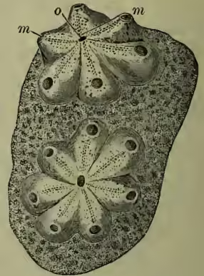
Rosettes of 6 or 7 united individuals, with separate inhalant, but united exhalant apertures; m, the inhalant aperture; o, the common exhalant aperture. The colonics are attached to a piece of seaweed.
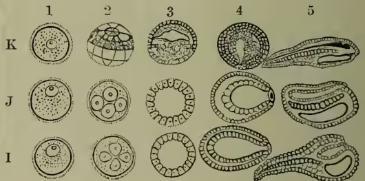
Development of: I, Ascidian; J, Amphioxus; K, Frog.
1, ovum; 2, segmenting ovum; 3, morula or blastosphere;
4, gastrula; 5, further advanced embryo. between the cavity of the canal and the original mouth of the gastrula. This communication, connecting nerve canal and primitive gut, is known as the neurenteric canal. On the back of the primitive gut, towards the posterior end, a band of cells is formed, the beginning of the notochord, which supports the growing tail, and represents in these lowly vertebrates the incipient 'backbone.' The sides of the primitive gut-cavity form posteriorly the lateral muscles. All this time the embryo has been inclosed within its egg-membrane, but with the growth of the tail, two or three days after fertilisation, the imprisoning case is burst, and there is liberated a free-swimming larva—strikingly like a tadpole. At the end of the closed canal, which may be said roughly to represent the spinal cord, there is a dilatation which may be called the brain. In this some progress soon becomes manifest, especially in the appearance of ear and eye, which remain in closest association with the brain, and are lost in the degenerate adult. The remains of the primitive gut form in various ways the respiratory pharynx, the oesophagus, stomach, and intestine of the adult ascidian.
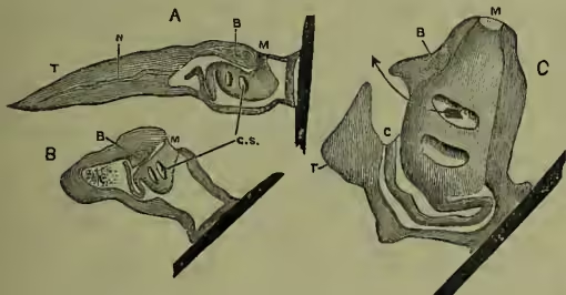
About the time the larva is hatched, another very important organ appears. A pair of outgrowths from the front of the gut meet, and fuse with a pair of dorsal involutions from the skin. The result is a paired atrial cavity, virtually open to the exterior by the two apertures of involution, and connected with the gut at the origin of the outgrowths—i.e. by gill-clefts. After a while a second pair of clefts appear, opening into the same atrial cavity. It must be noted at this stage that a secretion from the skin which began before the larva was hatched, has resulted in the formation of an intact 'test,' which clothes the entire larva, and is nowhere perforated. The free tadpole-like larva now fixes itself, first by a papilla and then by its test, as shown in the diagram, and with attachment the spinal cord, notochord, &c. begin to degenerate. An opening in the test appears opposite the mouth, which thns becomes at length functional. The paired atrial cavities also open by perforations through the test, and dilate internally so as to embrace the whole of the respiratory pharynx. To this their walls become, indeed, attached, and a large number of gill-slits perforate this double wall. The two openings to the exterior coalesce into one, and the two cavities also unite to form a single peribranchial chamber. The anus has been gradually shunted dorsally, and opens into the atrial cavity in the region already referred to as the cloaca. The gradual modification of structure is well illustrated in the accompanying figure. The result is in most cases a vivid instance of that degeneration which not unfrequently follows from sedentary life. See AMPHIOXUS, APPENDICULARIA, DEGENERATION, VERTEBRATA; also Huxley's Anatomy of Invertebrates; Balfour's Embryology, vol. ii.; Ray Lankester's Degeneration (in the Nature Series).