Brain is the term applied to that part of the central nervous system which in vertebrated animals is contained within the cranium or skull, and, in the invertebrata, to the nervous ganglia near the head end of the body. The brain is enveloped by three membranes, named the Dura-mater, the
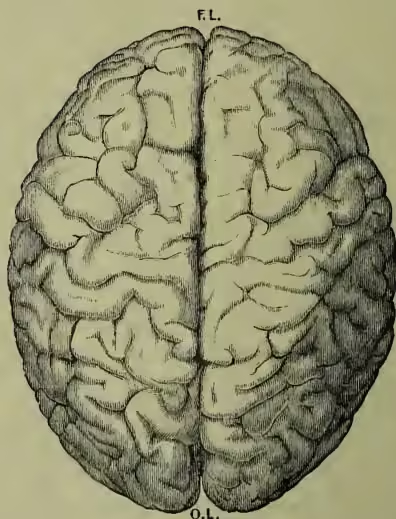
To show, firstly, division into two nearly equal hemispheres by the great median fissure; secondly, general appearance and apparent irregularity of arrangement of the convolutions and fissures; F.L., frontal lobe; O.L., occipital lobe.
Araehnoid, and the Pia-mater. These support the blood-vessels which nourish the cranium and
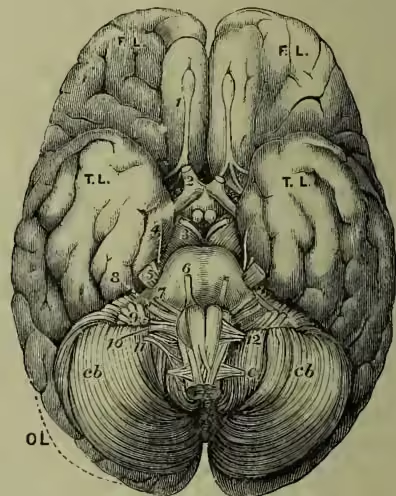
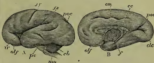
F.L., T.L., O.L., frontal, temporal, and occipital lobes of the cerebrum; cb, cb, cerebellum, the medulla oblongata lying between its two lobes. Cranial Nerves.—1, olfactory lobe (the nerve of smell); 2, optic nerve (nerve of sight); 3, third or oculo-motor nerve, motor nerve to most of the muscles of the eye; 4, fourth or trochlear nerve, motor nerve to the superior oblique muscle of the eye; 5, fifth, trigeminus, or trifacial, sensory and motor, the large root sensory to the face and eyes, &c.; the small root, motor to muscles of mastication; 6, sixth or abduces nerve, to external rectus muscle of eye, turns eyeball outwards; 7, seventh or facial, motor to muscles of expression; 8, eighth or auditory nerve, sensory for hearing (cochlea) and for equilibration (semicircular canals); 9, glosso-pharyngeal, sensory nerve of taste, and motor to some of the muscles of deglutition; 10, pneumogastric, sensory and motor to larynx, lung, heart, and stomach; 11, spinal accessory, motor to muscles of heart (inhibitory) and sterno-mastoid and trapezius; 12, hypoglossal, motor to all the muscles of the tongue; c1, first cervical spinal nerve. the brain, and also contain a clear fluid—the cerebro-spinal fluid—which removes the products of brain waste, and at the same time serves, like a water-cushion, to diminish the effect of external shocks. Brain substance is composed of gray and white matter (with some connective tissue and blood-vessels). The gray matter consists of nerve cells, which are minute structures, variously shaped, communicating by numerous fine processes with nerve fibres. These cells discharge nerve impulses to, and receive impressions from nerve fibres. The white matter is composed nearly entirely of white medullated nerve fibres, whose function is to transmit nerve impulses from one point to another. The relations of the gray and white matter vary greatly in the different parts of the brain. The main divisions of the brain are the cerebrum (great brain or cerebral hemispheres), the cerebellum (with the pons Varolii), the optic lobes (corpora quadrigemina, with the crura cerebri), and the medulla oblongata. An examination of figs. 1, 2, 3, 5, from different aspects of the human brain, will convey an idea of the relative size and position of these parts. The cerebrum underlies the whole vault of the cranium, and covers all the rest of the brain when seen from above (fig. 1). Below its posterior extremity, and separated from it by a fold of dura-mater, is the cerebellum. Below the cerebellum, again, lies the medulla oblongata, which is continuous, through an opening in the base of the skull called the foramen magnum, with the fissure, at the bottom of which lies a great transverse commissure, the corpus callosum. The hemispheres are covered with a thin layer of gray matter (or nerve cells), and thrown into ridges and furrows (technically called convolutions or gyri, and fissures). These seem (fig. 1) to be arranged without any definite order, but Gratiolet, by comparing the simpler brains of monkeys and of the human embryo, discovered that certain fissures ('primary' fissures) were fs, fissure of Sylvius; fr, fissure of Rolando; poc, parieto-occipital fissure; ec, calcarine fissure; em, calloso-marginal fissure; ir, island of Reil (not visible from the surface in the adult); olf, olfactory lobe; f, fornix; pv, pons Varolii; mo, medulla oblongata; cb, cerebellum.
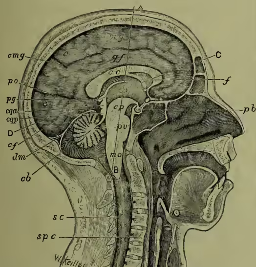
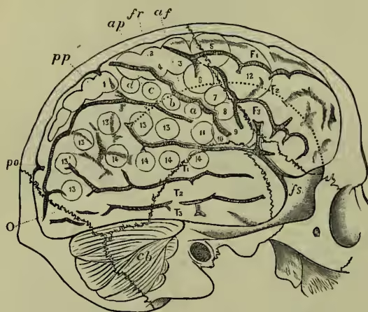
Spinal Cord (q.v.). The medulla passes directly upwards into the protuberance called the pons Varolii, which is connected by the crura cerebri or cerebral peduncles with the cerebrum. Behind the crura cerebri, in the angle between the cerebrum and cerebellum, lie the optic lobes (corpora quadrigemina). The cerebrum consists of two nearly equal hemispheres, divided by a deep median always present. There are, on the outer surface, the fissure of Sylvius (fs), the fissure of Rolando (fr), and, on the inner surface, the parieto-occipital (po). These fissures form the boundaries of the various lobes of the cerebrum. (1) The frontal lobe (FL) is that part of the outer surface, and the corresponding part of the median surface, which lies anterior to the fissure of Rolando. (2) The temporo-sphenoidal lobe (TL) lies below the fissure of Sylvius. (3) The occipital lobe lies behind the parieto-occipital fissure, and includes the corresponding parts on the outer surface. (4) The parietal lobe is bounded by the fissures of Rolando and of Sylvius, and by the occipital lobe. (5) The island of Reil lies at the bottom of the fissure of Sylvius (ir, fig. 4), and is obscured in the adult by the adjacent lobes. Secondary fissures on these lobes divide them into convolutions. Thus (fig. 5), on the frontal lobe, we have the first (F1), second (F2), third (F3), and ascending (af) frontal convolutions; on the temporo-sphenoidal, the first (T1), second (T2), and third (T3) temporal convolutions; on the parietal, the ascending (ap), postero-parietal (pp), angular (13'), and supra-marginal (13) convolutions. On the median surface (fig. 3) we have the marginal convolution, the gyrus fornicatus (gf), immediately above the corpus callosum, and the gyrus hippocampi, &c. On the under surface of the cerebrum we see the two olfactory nerves (1) and the two optic nerves (2). The latter, crossing like the letter X, wind round the two cerebral peduncles (cp), to terminate in the optic thalami and optic lobes. These peduncles are seen passing from the under surface of the hemispheres, and approaching each other as they enter the pons Varolii. If we press apart the two cerebral hemispheres, we come upon the corpus callosum (cc, fig. 3). This is a band of white fibres that probably connects corresponding convolutions of both hemispheres. On dividing this and removing some white fibres (the fornix, fig. 3, f), and a layer of connective tissue (velum interpositum), with its vascular margin (the choroid plexus), we expose the ventricles of the cerebrum—viz. the two lateral and the third ventricles. The former occupy the hemispheres, the latter lies between them, and is continued backwards through a narrow channel (the aqueduct of Sylvius) into the fourth ventricle, which lies behind the pons Varolii and medulla (figs. 6, 7, 8). Projecting into the third and lateral ventricles are rounded masses of gray matter, the corpus striatum and optic thalamus, often called the basal ganglia (figs. 6, 7). A better idea of the fr, fissure of Rolando; fs, fissure of Sylvius; po, parieto-occipital fissure; F1 superior, F2 middle, F3 inferior frontal; af, ascending frontal convolution; ap, ascending parietal; pp, postero-parietal convolution; T1 superior, T2 middle, T3 inferior temporo-sphenoidal convolution; O, occipital lobe; cb, cerebellum; 1 (postero-parietal convolution), advance of the opposite leg, as in walking; 2, 3, 4 (round upper extremity of fissure of Rolando), complex movements of opposite leg, arm, and of the trunk, as in swimming; a, b, c, d (ascending parietal convolution), individual and combined movements of the fingers and wrist of the opposite hand, or prehensile movements; forward extension of opposite arm and hand; 5 (posterior end of superior frontal convolution), forward extension of opposite arm and hand; 6 (upper part of ascending frontal convolution), supination and flexion of opposite forearm; 7 (median part of ascending frontal convolution), retraction and elevation of opposite angle of the mouth; 8 (lower part of ascending frontal convolution), elevation of ala nasi, and upper lip, with depression of lower lip; 9 and 10, opening of mouth with protrusion and retraction of tongue—on the left side is aphasic region; 11, between 10 and lower end of ascending parietal convolution, retraction of opposite angle of the mouth, the head turns slightly to one side; 12, posterior part of the superior and middle frontal convolutions (eyes open widely, pupils dilate, head and eyes turn towards opposite side); 13, 13' (supra-marginal and angular gyri), eyes move towards opposite side, or upwards and downwards (centre for vision); 14, superior temporo-sphenoidal convolution (centre for hearing). relation of all these parts will be gained by studying the two transverse sections (figs. 6 and 7) made in the direction of the dotted lines AB and CD in fig. 3. Surrounding the whole cerebrum is the thin convoluted envelope of gray matter, about a quarter inch thick. Within this lies the centrum ovale composed of white fibres. Connecting the two hemispheres is the corpus callosum (cc) with, underneath it, the septa lucida and fornix (fig. 7). Projecting into the ventricles are the ovoid optic thalamus and the caudate nucleus (cn) of the corpus striatum. A wedge-shaped mass of gray matter (ln), the lenticular nucleus of the corpus striatum, is separated from the first two nuclei by a band of white fibres, the internal capsule (ic), which in fig. 6 we see to be composed of two parts, an anterior limb and a posterior limb, which meet each other at an obtuse angle (the knee). Immediately outside the lenticular nucleus is the white external capsule (cc), separated by a thin band of gray matter, the claustrum, from the island of Reil (ir). The centrum ovale consists of nerve fibres passing in various directions, very difficult to unravel from each other. Certain of them pass between adjacent convolutions, others again connect parts at a distance, such as the frontal and occipital lobes, many cross by the corpus callosum to end probably in corresponding convolutions of the opposite hemisphere. Others connect the basal ganglia with the cortical gray matter. From the internal capsule an important group, known as the corona radiata (see fig. 7), passes to the whole of the cortex. That part of the corona radiata entering the occipital lobes is called the optic radiation of Gratiolet, who considered it to be the central expansion of the optic nerve (or, fig. 6).
The optic lobes consist of two (anterior and posterior) pairs of rounded eminences of gray matter (figs. 3 and 8) situated close to the optic thalami, and underlying the pineal gland. They are very intimately connected with the optic nerves, part of these ending in the anterior pair; and also to the third and fourth nerves, whose nuclei of origin lie just underneath them in front of the aqueduct of Sylvius (3', fig. 8). The crura cerebri are formed of fibres passing down from the cerebrum to the pons, medulla, and cord (see fig. 3), and of others passing up from the medulla and cord, and from the cerebellum (superior peduncles) to the cerebrum.
The cerebellum possesses a median and two lateral hemispheres which have been subdivided into lobes (figs. 2 and 3). Its parts are arranged in thin laminae or folia with deep intervening fissures.
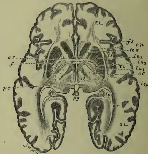
F.L., frontal; T.L., temporal; O.L., occipital lobes; fs, fissure of Sylvius; ir, island of Reil; cn, caudate nucleus; ln1, ln2, ln3, the three divisions of the lenticular nucleus; ica, anterior limb of internal capsule; icp, posterior limb of internal capsule (the anterior part conveys motor, the posterior sensory fibres), the part generally injured in cerebral hemorrhage; ot, optic thalamus; or, optic radiation, probably conveys sensory fibres to occipital and temporal lobes; ac, anterior commissure, connects both temporal lobes; f, fornix; pc, posterior commissure; pg, pineal gland; lv, lateral ventricle (posterior horn).
These laminae have a central core of white matter with a thin covering of gray matter. Sections through them recall the appearance of a tree and its branches (hence the term arbor vitæ). The cerebellum has three pairs of peduncles (fig. 8)—(a) superior, which pass upwards and across the middle line towards the opposite cerebral hemisphere, but ending under the optic lobes; (b) middle, which form the main part of the pons Varolii, and which enter indirectly into connection with fibres from the opposite cerebral hemisphere; (e) inferior (the restiform body), which is connected with the medulla and Spinal Cord (q.v.). There is also an intimate connection by the auditory nerve with the semicircular canals of the ear.
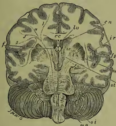
The shaded outline of the cerebrum indicates the superficial gray matter. fs, fissure of Sylvius; ir, island of Reil, a convolution at the bottom of the fissure of Sylvius concealed in the adult; cc, corpus callosum, the transverse interhemispherical commissure; lv, lateral ventricle; f, fornix, divided transversely. Between the fornix and the corpus callosum are seen the thin septa lucida; cn, caudate nucleus; ln, lenticular nucleus (with its three divisions); ot, optic thalamus; ie, internal capsule; ec, external capsule, and outside it a layer of gray matter, the claustrum; pv, pons Varolii; mo, medulla oblongata; oi, inferior olivary body; cb, cerebellum; l, a, f, s, mark the origin of the dotted lines representing motor fibres passing from the leg, arm, face, and speech 'centres,' through the internal capsule, cerebral peduncles, pons Varolii, medulla oblongata, and crossing at dp, the decussation of the pyramids to the opposite side of the spinal cord (to explain crossed paralysis). Another dotted line passes from s (speech centre) to hearing centre in first temporal convolution.
The Medulla Oblongata is the lowest division of the brain. It has a somewhat conical form, with the base towards the pons Varolii, the narrow end towards the spinal cord. On the under surface, on each side of the middle line, are the anterior pyramids decussating with (crossing) each other where the medulla passes into the spinal cord. External to them is an ovoid projection, the olivary body (fig. 2). On the dorsal aspect is the lozenge-shaped fourth ventricle, which is bounded below by the two inferior and above by the superior peduncles of the cerebellum. All the cranial nerves below the fourth originate from the floor of the medulla in the positions marked by the numbers on the right side of fig. 8. Their points of emergence on the under surface are indicated in fig. 2.
SIZE OF BRAIN.—The brain of the male has an average weight of 49½ ounces, that of the female is about 5 ounces lighter. Cuvier's brain weighed 1861 grammes (1 gramme is 15½ grains), Turgeneff's 2012 grammes. On the other hand, Virchow has found a brain weighing 1911 grammes in a man without any specially high mental development, and the brains of some very able men have been found below the average weight. Too much stress has been laid upon the connection of mental capacity with brain weight in individual instances. Still the general fact is that the most highly developed races have the heaviest brains.
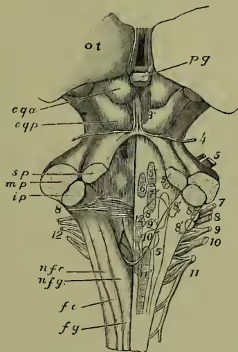
FUNCTIONS OF THE CEREBRUM.—If we remove the cerebral lobes from an animal, we deprive it of its volition and its intelligence. We may leave it capable of performing very complex movements, almost, if not quite, as perfectly as before; but we have destroyed its power to initiate these movements, we reduce it to the condition of a responsive machine. Thus a frog without its cerebrum will, if undisturbed, remain motionless for an indefinite time. If it be placed on a board, and the board tilted, it will change its position till its equilibrium is stable, and then it becomes motionless. If put into water, it will swim, and if a piece of wood be put before it, it will climb up on it, and there remain; if it be pinched, it will spring and will avoid any obstacle placed in its way, just as an entire frog would. A pigeon similarly treated becomes drowsy and stupid. If food be placed before it, it will not take it; it exhibits no sign of terror at what would otherwise cause it alarm, further than to start slightly at a loud sound, or a bright light. But if thrown into the air, it will fly to a convenient resting-place; if placed on its back, it will regain its feet, and thereafter become drowsy again. ot, optic thalamus; pg, pineal gland; cqa, anterior corpora quadrigemina; cqp, posterior corpora quadrigemina; sp, superior cerebellar peduncle; mp, middle cerebellar peduncle, goes to pons Varolii; ip, inferior peduncle, or restiform body, goes to medulla oblongata and spinal cord; fy, funiculus gracilis, or column of Goll; nfg, nucleus of the funiculus gracilis; fc, funiculus cuneatus, or column of Burdach; nfc, nucleus of the funiculus cuneatus. The lozenge-shaped area in the centre of the figure is the fourth ventricle. The numbers 4-12 indicate the superficial roots of the corresponding cranial nerves. The numbers 3'-12', their nuclei of origin.
Similar experiments on other animals show similar results. In man we find that imperfect development of the cerebrum is accompanied by imbecility and idiocy, and that the races that have the heaviest cerebrum and the most fully developed convolutions are the most intelligent. Among animals, again, the degree of intelligence increases with the increase in size of the cerebrum relatively to the other parts of the brain.
Flourens, the great French physiologist, held that the whole of the cerebrum was employed in every mental process. He removed parts of the hemispheres of pigeons, and came to the conclusion that all the mental functions became enfeebled, and that to an equal degree, in proportion to the amount of brain matter removed, no matter from what part of the cerebrum it were; and that as long as any part of the gray matter remained, so long could mental functions be carried on. This doctrine long held the field. It seemed to explain why large masses of brain substance could be removed by injury without apparent effect on the mind, and why there was no constant relation between symptoms and locality of brain lesions. But it did not explain all the facts. The condition of Aphasia (q.v.), or loss of articulate speech, was found to be almost constantly associated with a certain convolution, and certain paralysis and limited convulsive phenomena were found by Hughlings Jackson to be apparently due to definitely localised lesions. In 1870 Fritsch and Hitzig, two German observers, discovered that when certain areas of the cerebral cortex were stimulated by the galvanic current, movements of the opposite side of the body were produced. This discovery put the question of cerebral localisation on a new footing. Ferrier repeated and extended their results, and soon succeeded in mapping on the brains of animals (rabbits, dogs, and monkeys), areas, stimulation of which by the faradic current produced movements which corresponded to the area as accurately as the notes correspond to the keys of a pianoforte. A great controversy immediately arose as to the meaning and value of these discoveries, the result of which has been to substantiate their accuracy, and to lead to their extension. Ferrier's 'motor areas' (see fig. 5) lie on either side of the fissure of Rolando, and extend into the postero-parietal and part of the first, second, and third frontal convolutions. The lower part of the area corresponds to the movements of the face and tongue; the intermediate parts to those of the arm; the upper to those of the leg. Recently, a centre for the muscles of the trunk, arm, and leg has been found on the marginal convolution (on the median aspect of the parietal lobe); and Ferrier's areas have been further subdivided, so that centres for such fine movements as, e.g., the apposition of the forefinger to the thumb have been mapped out on the brain of the monkey by Horsley, Schäfer, and others. A knowledge of the correspondence between the convolutions of the simian and the human brain, and of the relation of the convolutions of the latter to the bones of the skull (see fig. 5), has already led to the successful removal of tumours whose seat was indicated by definite motor disturbances. When these areas are destroyed by disease, the power of the corresponding voluntary movement or movements is lost. When they are irritated, corresponding spasms of movement are set up (Jacksonian epilepsy).
Aphasia.—The left hemisphere controls the right side of the body, and as we are mostly right-handed, so we are left-brained. When the posterior end of the third frontal convolution on the left side is destroyed, aphasia results. An aphasic person understands what is said to him, but has lost the power of articulate speech—the words wanted are either forgotten, or cannot be uttered, and this although the muscles of articulation are unimpaired. They cannot be co-ordinated. In left-handed individuals aphasia has been found associated with the corresponding lesion on the right side of the cerebrum. The third left frontal convolution is said to be well developed in the brains of remarkable men, and to be very simple in deaf-mutes, and quite rudimentary in idiots. The corresponding condition of agraphia, or inability to express one's self intelligently by writing, has been recently found associated with lesions of the posterior end of the second frontal convolution.
From these various areas white nerve fibres pass downwards through the centrum ovale, and converge toward the internal capsule, where they occupy the anterior two-thirds of the posterior limb, the fibres from the face and tongue being farthest forward, then come those for the arm, and lastly those for the leg. The fibres that go to the nuclei of the facial and tongue muscles, cross the middle line a little below this, those for the limbs and arm at the decussation of the pyramids at the lower end of the medulla. When a cerebral hemorrhage injures them in the internal capsule (the most frequent seat), crossed paralysis will follow, with aphasia if the lesion has been on the left side (fig. 7, l, f, a, s).
Cortical Sensory Areas.—The determination of the existence of definite areas for the conscious perception of the different forms of sensory impressions is much more difficult than that of the motor areas, and physiologists are yet far from being agreed on this point.
Centre for Sight.—Munk places this centre in the occipital lobe; destruction of one lobe produces permanent blindness in one side of both eyes (what is called homonymous hemianopsia). Ferrier destroyed the occipital lobes in monkeys, and found no appreciable alteration of vision, but when in addition both angular gyri were destroyed, permanent blindness followed. Destruction of one angular gyrus caused only temporary loss of vision in the opposite eye, while destruction of both caused a similar temporary condition in both eyes. In man, disease of the left angular gyrus produces what is called word-blindness. In this condition one loses the power of reading words, although one sees the characters distinctly, and may even be able to spell the word. One may even retain the power of writing spontaneously or from dictation, and yet be unable to read what has been written. It may be compared to the condition where one has completely forgotten a foreign language; one can write or read the words, but has absolutely lost their meaning. Recent investigation seems to show that the occipital lobes alone are the centre for sight, each lobe being connected with the corresponding lateral halves of the two eyes.
Centre of Hearing.—This seems to lie in the first tempo-sphenoidal convolution in both sides. In man, partial destruction of this convolution on the left side causes the condition of word-deafness (where one hears the sounds, but has absolutely lost their meaning).
Centre for Taste and Smell.—Ferrier locates the former of these in the uncinate gyrus (on the inner surface of the tempo-sphenoidal lobe).
Centre for Touch (common sensibility).—Many physiologists place these in the motor area. Ferrier, Horsley, and Schäfer find that they are in the gyrus fornicatus and the gyrus hippocampi (on the median aspect of the brain, above and behind and below the corpus callosum, fig. 1).
Fibres from the various sense organs lead to the cortical areas, through the posterior third of the internal capsule. If this is injured, loss of all forms of sensibility—hearing, sight, taste, touch, &c.—of the opposite side follows.
Frontal Lobes.—No definite result follows from the experiments made on the anterior part of the frontal lobes; but it is probable that they are associated with the exercise of the higher mental faculties. They are well developed in highly intelligent individuals, and vice versa. It must be admitted, however, that cases have been recorded (as in the American case of a man injured in this region by a crowbar) where the frontal lobes have been greatly damaged without apparent effect on the mind. (It is usual to speak of the cerebrum as being the seat of the various mental faculties, and of the will as producing voluntary movement. All we know is this—that changes in consciousness are accompanied by molecular changes in the brain; the one does not produce the other. So it is nervous activity that produces movement; not the mental action of the will.)
Basal Ganglia.—Stimulation of the corpus striatum causes general contraction of the muscles on the opposite side of the body, as if the whole motor area were being stimulated at once. Destruction of the optic thalamus seems to cause interference with the vision (the optic tract ends partly in its posterior part), and sometimes with other forms of sensation. Further investigation, however, is needed to establish definitely the functions of these ganglia.
Optic Lobes.—The optic lobes seem to be concerned in the co-ordination of those movements noted in the frog as persisting after removal of the cerebral hemispheres. A frog without these lobes loses the power of recovery from a disturbance of its equilibrium, or of properly performing any other set of combined movements. In man the anterior pair of the corpora quadrigemina seems to control the reflex contraction of the pupils, and of some of the movements of the eyes through the third nerve; while the posterior pair has some more general co-ordinating power. Unilateral lesions cause in animals what are called 'forced movements'—i.e. a tendency to run in a circle, like a horse in a circus, or to turn like the hand of a watch round the tip of the tail as a centre, or to rotate round the axis of the body.
Cerebellum.—Flourens considered that the function of the cerebellum was to co-ordinate voluntary movements. He found that removal of superficial layers in a pigeon caused slight weakness and disharmony of movement; that further removal caused general agitation, and finally loss of ability to stand or fly or walk. It could not rise when placed on its back. But it was not paralysed, it made continual ineffectual struggles to recover its position, and only ceased when completely exhausted. There was no loss of volition or intelligence. It could see and hear and feel quite well. Disease of the human cerebellum, however, when it causes symptoms, does not cause general incoordination, but merely a staggering gait like that of intoxication, and a feeling of giddiness. Ferrier holds that it is the centre for co-ordinating movements necessary for the equilibrium of the body, and that its different parts control different sets of these movements. For instance, destruction of the anterior part of the median lobe causes tendency to fall forwards, loss of its posterior part causes tendency to fall backwards, and of one lateral lobe a tendency to rotate towards the side injured. Stimulation of any one of these parts causes movements of the head, eyes, and limbs, such as would counteract the disturbance of equilibrium produced by the destruction of the part. The anatomical connections of the cerebellum, with the semicircular canals of the ear, seem to point to the same conclusion. Other observers have regarded the cerebellum as the seat of the muscular sense, or for the maintenance of muscular tone, but without sufficient warrant.
The Medulla Oblongata is the great seat of the centres for the functions of organic life, as may be understood from the fact that it gives origin to all the cranial nerves, except the first four pairs (see fig. 8). The various centres are: (1) The centre for respiratory movements, near the lower extremity of the fourth ventricle; (2) for the inhibition (or restraint), and for the acceleration of the movements of the heart; (3) for the control of the blood pressure (vaso-motor centre), including the 'diabetic' centre, which is simply the vaso-motor centre for the liver; (4) centre for swallowing; (5) centre for movements of the gullet and stomach, and the vomiting centre; (6) for movements of articulate speech; (7) for the secretion of the saliva. The term 'centre' involves the following mechanism: (1) A sensitive end-organ or surface; (2) an afferent nerve going to (3) a nerve cell, or group of nerve cells, from which passes (4) an efferent nerve fibre to (5) a muscle. That the action of the 'centre' be possible all the links in the chain must be intact.
DISEASES OF THE BRAIN.—Abscess.—Abscess of the brain is a collection of pus in its substance. The most common causes are compound fractures of the skull, diseases of the middle ear and temporal bone, and pyæmia. Many cases develop quietly, others are preceded by headache, pain, vomiting, delirium. When pus has formed, the symptoms will depend on the site; usually there is more or less paralysis of motion. Finally the signs of compression precede the fatal termination. When an abscess in the brain has formed, if its situation can be made out, the pus should be evacuated by Trephining (q.v.). Many cases have recently been treated successfully in this way.
Anæmia of the brain is a deficiency in the quality or in the quantity of blood in the brain. It may result from direct loss of blood, exhausting diseases, weakness of the heart, or from the pressure of growths or fluids within the skull. Headache, drowsiness, hyperæsthesia, giddiness, muscular weakness are generally present, and most marked in the erect position. Syncope, or fainting, may result from the too rapid assumption of the erect position. In anæmia from acute loss of blood, and in syncope, the head must be kept low, and stimulants applied to the skin, and, if possible, administered internally. In chronic anæmia, the cause must be removed, stimulating nourishment given at frequent intervals, and iron administered.
Atrophy of the brain is a wasting of its substance, and is either congenital, or a condition accompanying old age, or resulting from exhausting disease, and very frequently from alcoholic excesses. The congenital form produces imbecility or idiocy. The acquired form leads to loss of memory and of mental power, and generally to diminution of muscular power.
Compression, concussion, and contusion of the brain are three terms often popularly confounded with each other. Compression means the squeezing of the brain by any fluid such as blood, serum, or inflammatory exudation or tumour. Concussion is the result of shock to the brain from an injury, but without visible effect on its substance. In contusion the brain substance is more or less lacerated. In compression there is paralysis, insensibility, coma gradually deepening, dilated pupils, and generally a slow pulse. In concussion there is first a period of collapse, with pallor of face, feebleness of pulse, and cold extremities. If this is recovered from, consciousness gradually returns with vomiting, fever, and a more or less sleepy condition. Perfect quiet, and the avoidance of all stimulation, must be observed till convalescence is complete.
Congestion of the brain causes headache, giddiness, mental excitement, and sleeplessness. If long continued, a period of depression follows with drowsiness, mental and physical weakness. Some attacks are so acute as to produce delirium, or convulsions, or even symptoms of apoplexy, which may be fatal. Among the causes of acute congestion may be noted excessive mental strain, the use of alcohol in excess, and some of the acute fevers. It is of frequent occurrence in certain diseases of the nervous system.
Inflammation of the brain substance is usually the result of injury or disease of the bones of the skull, and is accompanied with more or less meningitis.
Hemorrhage into the brain leads to Apoplexy (q.v.). It is probably always preceded by disease of the blood-vessels, more especially with chronic Bright's disease and alcoholism. The vessels which rupture are most commonly the small arteries which pass through the basal ganglia. The onset may be quite sudden, or there may be premonitory symptoms such as headache, giddiness, or numbness in the limbs, &c.
Meningitis, Hydrocephalus (or water in the head), Epilepsy, Aphasia, are all forms of brain disease, but are treated as separate articles.
Brain Fever is a popular term which includes congestion of the brain and its membranes, delirium tremens, and inflammation of the brain substance itself. It ought to be discontinued altogether.
Softening is a term popularly and very erroneously used to indicate failure of mental power and general feebleness, often accompanied with drowsiness, dullness, loss of memory, and with emotional excitability. There may be no actual softening in such a condition—merely atrophy of the brain.
True softening is the result either of clotting of blood in the vessels, disease of the walls of the arteries (atheroma), or Embolism (q.v.), or the presence of tumours. The brain substance becomes red, or yellow, or white, and the fibres break up as oil drops, and are gradually absorbed, a cyst containing fluid being left. The symptoms will depend on the part affected; if in the motor area, there will be paralysis of motion; or in the sensory area, loss of the corresponding power of conscious perception.
Tumours may grow from the membranes, the blood-vessels, or the connective tissue of the brain. They vary greatly in structure, many being allied to the sarcomata. The symptoms vary greatly according to their position, or may be absent entirely. The most common symptoms are intolerable headache, vomiting (without sickness), giddiness, convulsions, paralysis, and a condition of the optic nerve and retina recognisable by the ophthalmoscope, called optic neuritis. Some tumours can be removed by medical treatment, a few by surgical means. Generally all that can be done is to relieve pain and support the strength.
See also the articles ANIMAL MAGNETISM, APHASIA, APOPLEXY, CEREBRO-SPINAL FLUID, CONCUSSION, CONSCIOUSNESS, EPILEPSY, HYDROCEPHALUS, Hysteria, INSANITY, INSOMNIA, MEMORY (DISEASES OF), MENINGITIS, NERVOUS DISEASES, NERVOUS SYSTEM, PARALYSIS, PERSONALITY, RIGHT-HANDEDNESS, SHOCK, SOMNAMBULISM, SUNSTROKE; Ferrier, Functions of the Brain (2d ed. 1886); Calderwood, Mind and Brain (1879); Quain's Anatomy (8th ed.); Ross's Diseases of the Nervous System (2d ed. 1883); and papers by Horsley, Schäfer, and others, in the Philosophical Transactions.