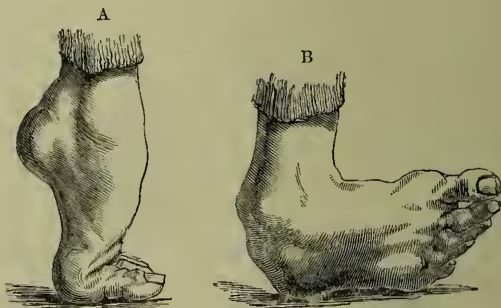
Club-foot (Lat. talipes) is a distortion of the foot primarily due to shortening occurring in one or other of the groups of muscles which carry out its intricate movements; subsequently, this error in the muscular activity becomes aggravated by shortening of the ligaments which bind the bones together, and ultimately the shape of the bones themselves becomes altered so as to constitute a very serious deformity, difficult to correct. In the majority of cases the condition is congenital, and at the time of birth it is usually only the muscular structures that are affected. Thus it is of great importance to recognise its presence early in life; for until the ligaments and bones become altered, it may be remedied by very simple means, such as manipulation and electric stimulation of the affected groups of muscles. If these simple measures appear ineffectual, recourse must be had to the division of the tendons (Tenotomy, q.v.), by which the shortened muscles are attached to the bones of the foot. The foot thus freed from the cause of the distortion, should be placed at rest in good position within an immovable apparatus, such as a plaster of Paris case, until healing of the tendons has occurred. The weakened parts should then be exercised by suitable manipulations which restore their strength. In very bad cases it may be necessary to remove portions of the distorted bones and thus restore the natural shape of the foot.
These affections are very markedly hereditary, and they are in all probability due to disordered function in the nerves leading to the affected muscles, or in the brain or spinal cord, in which these nerves have their origin. Four chief varieties of club-foot are recognised by surgeons: (1) Talipes equinus (fig. A), in which the heel is drawn up, and the patient walks on the under surfaces of the toes; (2) Talipes calcaneus, in which the reverse condition is present, and the patient walks on the heel only; (3) Talipes varus (fig. B), in which the patient walks on the outer border of the foot; and (4) Talipes valgus, where the inner edge of the sole alone touches the ground. See DEFORMITIES.