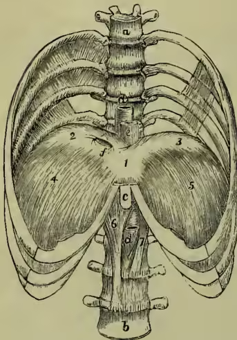
Diaphragm, or MIDRIFF (Gr. diaphragma, 'a partition'), is the name applied in anatomy to designate the musculo-tendinous partition which in man and the mammalia generally separates the cavity of the thorax from that of the abdomen. Its general form is that of a dome directed towards the chest, the lower part and sides being muscular, while the central or highest portion consists of an expanded tendon. The muscular fibres have an extensive origin from the circumference of the visceral cavity—viz. posteriorly, a vertebral portion arising from the lumbar vertebrae by two pointed processes, or crura, of unequal length, and from two fibrous bands, or arched ligaments, on each side of the bodies of the vertebrae: laterally, a costal portion arising from the cartilaginous and adjoining osseous parts of the lower six ribs; anteriorly, a sternal portion arising from the ensiform cartilage of the sternum, or breastbone. From these various points the muscular fibres converge to the central, trefoil, or cordiform tendon, which forms the highest part of the diaphragm, and as its name indicates, consists of three lobes. The diaphragm presents three large perforations or foramina; one, quadrilateral, placed in the highest part of the tendinous centre for the upward passage of the inferior vena cava; one, oval in form, surrounded by muscular fibres derived from the crura, for the passage of the oesophagus and pneumogastric nerves; the third, situated in front of the vertebrae and bounded by tendinous fibres of the crura, transmits the aorta, thoracic duct, and large azygos vein.
The upper surface of the diaphragm is in relation to the pleural membranes which inclose the lungs, and the pericardium which incloses the heart. The latter membrane is intimately connected with the central tendon, which thus forms a fixed floor for the support of the heart. The lower surface, deeply concave in form, is lined by peritoneum, and has in apposition with it the stomach and spleen on the left side, the convex upper surface of the liver on the right side, and the kidneys, supra-renal capsules, and duodenum posteriorly.
When the diaphragm contracts, the arched portion becomes flatter, and the cavity of the chest being thereby enlarged, air rushes in to fill the vacuum, and expands the lungs during the act of inspiration. It is thus an inspiratory muscle, and in ordinary quiet breathing the central tendon on which the heart rests takes no share in this movement. Every contraction of the diaphragm must diminish the abdominal space and compress the abdominal viscera, and hence it aids in the expulsion of the faeces and urine.
Spasmodic action of the diaphragm produces hicough and sobbing. Stoppage of the action of the diaphragm, whether from great external pressure or from paralysis, is very speedily fatal.
DIAPHRAGM, a partition with a hole in it, employed not only in landscape and portrait lenses for photography, but also in telescopes, microscopes, and other optical instruments, for the purpose of cutting off the superfluous rays of light, and producing greater intensity or sharpness of the image, as well as to correct aberration.