Epithelium is the term applied in anatomy to the cell-tissue which, in layers of various thickness, invests not only the outer surface of the body, and the mucous membranes connected with it—as, for example, those of the nose, lungs, intestinal canal, &c.—but also the closed cavities of the body, such
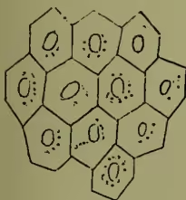
Epidermis (still soft like the epithelium of internal parts) of a two months' human embryo. Mag. 350 diam.
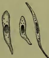
Epithelial cells of the vessels; the longer one from the arteries, the shorter ones from the veins.
as the great serous membranes, the ventricles of the brain, the synovial membranes of joints, the interior of the heart and of the blood-vessels proceeding to and from it, the ducts of glands, &c. The thickness of this tissue varies extremely with the position in which it occurs. In some parts it consists of numerous strata of cells, collectively forming a layer of more than a line in thickness; in other parts it is composed of only a few strata, or often of only a single stratum of cells, and can only be detected by the microscope. The cells of which the epithelium is composed are usually soft nucleated cells; they may be rounded, polygonal, fusiform, cylindrical, or conical in shape, and sometimes they possess vibratile cilia. No blood-vessels exist in epithelial tissues, although minute channels may be found between the cells, by means of which the plasma derived from sub-adjacent blood-vessels may pass for the nutrition of the cells. In many cases nerve fibrils are abundant.
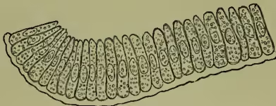
Epithelium of the intestinal villi of the rabbit. Mag. 300 diam.
Epithelia may be classified (a) according to the embryonic layer from which they are developed; (b) according to their function; (c) according to their shape and arrangement. Following the latter method, we have (a) simple epithelium, consisting of a single layer of cells which may be: (1) Pavement, consisting of polygonal plates or scales joined together by their edges. They constitute the variety known as pavement or tessellated epithelium, and occurring as an investment of the serous membranes, of most synovial membranes, of the lining membrane of the heart and of the veins, of the canals of glands, &c. (2) Columnar or cylinder epithelium, as in the intestine from the stomach to the termination of the alimentary canal, in the excretory ducts of all the glands opening into the intestine, &c. Illustrations of this cylinder epithelium are given in the article DIGESTION. (3) Spheroidal or glandular epithelium is chiefly characteristic of the terminal recesses of secreting glands. (4) Ciliated epithelium consists of cells which bear on their free ends spontaneously moving filaments called Cilia (q.v.). (b) Stratified epithelium consists of cells arranged in many layers, and the individual cells present every variety of shape. As a rule the deepest cells are columnar, and those on the surface flattened and scale-like, but overlapping one another at their margins. It occurs on the anterior
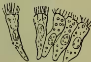
Ciliated cells from the finer bronchial tubes. Mag. 350 diam.
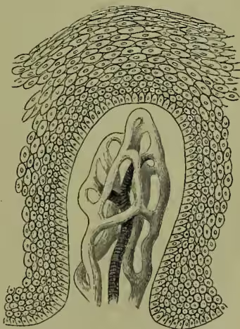
Stratified pavement epithelium investing a simple papilla (with blood-vessels in the interior) from the gums of a child. Mag. 250 diam.
surface of the cornea of the eye, in the mouth, pharynx, œsophagus, &c., but its most extensive distribution is in the Epidermis (q.v.). (c) Transitional epithelium is intermediate between the forms already described, and may be grouped under the three terms columnar, ciliated, and scaly transitional, according to the kind of cell which is most superficial. In the case of the columnar and ciliated varieties there are smaller cells irregularly disposed between the fixed ends of the large ones, and this constitutes the only difference between these and the columnar and ciliated cells already described.
Scaly transitional epithelium is found lining the urinary Bladder (q.v.) and ureters. The superficial cells are flattened scales when the bladder is distended, but cubical when empty. Moreover, the free surface of the cells is smooth, but on the deep aspect they are moulded over the rounded ends of the pear-shaped cells beneath. Irregular cells fill up the intervals between the tapering ends of the pyriform cells.
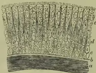
Ciliated epithelium from the trachea of a man. Mag. 350 diam. a, outermost part of the elastic longitudinal fibres; b, homogeneous outermost layer of the mucous membrane; c, deepest round cells; d, median long cells; e, outermost conical ciliated cells.
In all the varieties of epithelium the layer of external cells is being constantly disintegrated and replaced by the layer immediately beneath. The polygonal or pavement epithelium mainly acts like the epidermis, as a protecting medium to the soft parts beneath. The cylindrical epithelium additionally takes an active part in the process of secretion. Illustrations of the function of the cells forming this variety of epithelium are given in the articles CELL, CILIA, DIGESTION; and the subject will be further noticed under the head SECRETION.