Fœtus, the term applied in Medicine to the mammalian embryo, especially in its more advanced stages. In the human subject we usually speak of the embryo at and after the end of the fourth month as a fœtus.
The weight and length of the fœtus at full term—nine months—varies within considerable limits. Observations on a very large number of cases have shown that this variety is related to several conditions—e.g. sex, race, number of previous pregnancies of the mother, &c. Male children are from 8 to 12 oz. heavier than female; children born in Britain are heavier by 3 or 4 oz. than those born in France, and first-born children are 4 to 6 oz. lighter than subsequent ones.
On an average the fœtus at birth weighs from 6½ to 9 lb. But a healthy child may weigh as little as 5 or 5½ lb., and cases are on record where the child has weighed 17½ and 18 lb. Children under 5 lb. weight at birth rarely live, and when they do are puny. The length of the fœtus is 17 to 21 inches, occasionally being as much as 24 inches.
There are certain points in which the fœtus at the full period differs anatomically from the child shortly after birth. The bony skeleton is very incomplete, cartilage occurring in the place of many bones. Indeed, complete ossification (viz. of the vertebræ) is not finished until about the twenty-fifth year, and the only bones completely ossified at birth are the minute ossicles of the ear. The difference between the fœtus and the child in this respect is, however, only one of degree.
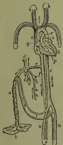
During pregnancy a temporary organ, termed the placenta (popularly known as the after-birth, from its being thrown off shortly after the birth of the child), is developed on the inner wall of the uterus (see b in the figure). This organ is mainly composed of vessels, and there proceeds from it the structure known as the umbilical cord, a, in which lie the umbilical vein, which conveys arterial blood to the fœtus, and the two umbilical arteries, which return the blood to the placenta. This umbilical cord conveys these vessels to the umbilicus or navel. Before tracing the course of the blood through the fœtus, we must notice the chief anatomical peculiarities presented by the vascular or circulating system before birth.
(1) In the heart we find a communication between the two auricles by means of an opening termed the foramen ovale. (2) In the arterial system we have to notice first the ductus arteriosus (see r in the figure), which is a large communicating trunk between the pulmonary artery, q, and the descending aorta, s; and, secondly, the branches given off by the internal iliac arteries, which go under the name of hypogastric as long as they are within the body of the fœtus, and of umbilical when they enter into the structure of the cord, are continued from the fœtus to the placenta, to which they return the blood which has circulated in the fœtal system. (3) In the venous system there is a communication between the umbilical vein, c, and the inferior vena cava, f, called the ductus venosus, e.
Pure blood is brought from the placenta by the umbilical vein, which passes through the umbilicus, and enters the liver, where it divides into several branches, d, d, which are distributed to that viscus, the main trunk or ductus venosus, e, passing directly backwards, and entering the inferior vena cava, f. The pure blood here becomes mixed with the impure blood which is returned from the lower extremities and abdominal viscera, and is carried into the right auricle, l, and from thence, guided by the Eustachian valve (which is situated between the anterior margin of the inferior vena cava and the auriculo-ventricular orifice, and is of relatively large size in the fœtus), passes through the foramen ovale, into the left auricle, i. From the left auricle it passes into the left ventricle, k, and into the aorta, l, whence it is distributed by the carotid and subclavian arteries principally to the head and upper extremities, which thus receive comparatively pure blood. From the head and arms the impure blood is returned by the superior vena cava, o, to the right auricle; from the right auricle it is propelled, as in the adult, into the right ventricle, p; and from the right ventricle into the pulmonary artery. In the adult it would now pass through the lungs, and be oxygenised; but in the fœtus it passes through the ductus arteriosus into the commencement of the descending aorta, where it mixes with that portion of the pure blood which is not sent through the carotid and subclavian arteries. Some of this mixed blood is distributed by the external iliac arteries, u, u, to the lower extremities, while the remainder (probably the larger portion) is conveyed by the hypogastric or umbilical arteries, t, to the placenta.
From the above description we perceive: (1) That a considerable quantity of the pure blood from the placenta is at once distributed to the liver, which accounts for its large size at birth as compared with the other viscera. (2) That a double current meets in the right auricle, one stream, guided by the Eustachian valve, passing through the foramen ovale into the left auricle, the other through the auriculo-ventricular opening into the right ventricle. (3) That the comparatively pure blood sent to the head and arms, as contrasted with the impure blood sent to the lower extremities, causes the relatively greater development of the former organs, and prepares them for the functions they are called upon to perform; the development of the legs at birth being slight as compared with that of the head or arms.
Almost immediately after birth the foramen ovale becomes closed by a membranous layer, and the ductus arteriosus and ductus venosus degenerate into impervious fibrous cords. The lungs, previously to the act of inspiration, are dense and solid in structure, and of a deep-red colour, and lie far back in the chest. Their specific gravity is greater than water, in which they (or portions of them) consequently sink, whereas lungs (or portions of lungs) that have respired float in that fluid.
Although nine months is the normal period of fœtal development in utero, it is possible for a fœtus to survive though born much within this period. When a child is born in a state of development sufficient to enable it to survive, it is said to be viable. A fœtus born at 4½ months may give evidence of life, such as movement of limbs, attempts at respiration, &c., but of course cannot survive, and hence is not viable. Several instances have been recorded in which the fœtus survived after birth at 6½ months. At seven months viability is established, and with reasonable care a very large proportion of children born at this time survive. Such children may become perfectly healthy and strong, both mentally and physically. It is said that Sir Isaac Newton was born at the seventh month. The French code fixes the longest limit of gestation at 300 days—a limit rarely reached, if ever.
This article would be imperfect without a notice of the question—What constitutes live-birth? This is a point on which the most distinguished obstetric authorities have differed: some holding that where there is muscular movement there is life; while others maintain that where respiration has not been proved to have taken place the child was still-born. Amongst the most celebrated lawsuits bearing on this point we may mention that of
Brook v. Kellock, tried in 1861, in which it was decided that a child may live for some time after birth and not breathe, the absence of signs of breathing being held to be no proof of its being born dead. It was given in evidence that there was pulsation of the funis after separation of the cord, and the beating of the heart was regarded as proof of live-birth. Hence we may regard it as established in English law that respiration is not required to establish live-birth. Nor do the laws of France or the United States require that the child shall have breathed. In Scotland the law requires not only that the child shall have breathed, but that it shall have cried; and in conformity with this law a child which lived, breathed, and died in convulsions at the end of half an hour was declared to have been born dead. See EMBRYOLOGY, and other articles cited there; GESTATION, PREGNANCY.