Foot. In man the bones of the foot are twenty-six in number, and are arranged in three natural groups—viz. the tarsal bones, which are the hindmost; the metatarsal bones, which occupy the middle portion; and the phalanges of the toes anteriorly. The tarsal bones, seven in number, are short and somewhat cubical, and form the heel and the hinder part of the instep. The uppermost (see fig. 1) is called the astragalus, from its supposed resemblance to the dice used by the Romans. Above, it articulates with the two bones of the leg, the tibia and fibula, and through these bones the whole weight of the body is thrown upon the two astragali. Below, it is connected with and rests upon the os calcis, or heel-bone, which is the largest bone of the foot. Immediately in front of the astragalus, and supporting it in this direction, is the scaphoid or boat-like bone.
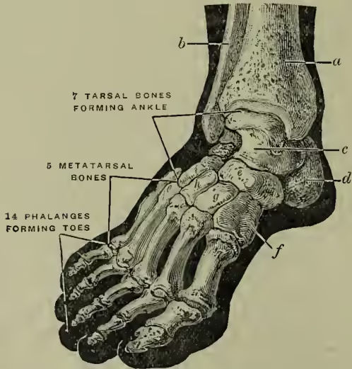
In front of the scaphoid bone are the three cuneiform or wedge-shaped bones; and on the outer side of the cuneiform bones, and in front of the os calcis, is the cuboid bone. We see from fig. 1 that the front row of tarsal bones is composed of the three cuneiform bones on the inner side of the foot, and of the cuboid bone externally. There are five metatarsal bones passing forward, one for each toe. Each cuneiform bone is connected with one, and the cuboid bone with two, of these metatarsal bones. Behind, they are close together, but as they run forward they diverge slightly from one another, and their anterior ends rest upon the ground and form the balls of the toes. They constitute the forepart of the instep. The remaining bones are those of the toes, and are named the phalanges, each toe having three of these bones, excepting the great toe, which has only two. (A similar law holds for the bones of the hand, each finger having three phalanges, but the thumb only two.)
The instep is composed of the seven tarsal and the five metatarsal bones, which are so arranged and connected (see fig. 2) as to form the plantar arch from the extremity of the heel-bone to the balls of the toes. The astragalus forms the summit or keystone of this arch, and transmits the weight which it receives back to the heel, and forward to the balls of the toes.
The bones where they articulate with one another are covered with a tolerably thick layer of smooth cartilage, and by this means, together with the very slight movements of which each bone is
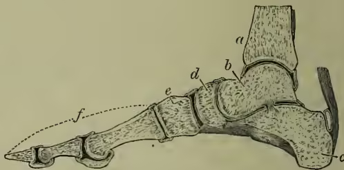
Section through the lower end of the tibia a, and through the astragalus b, the heel-bone c, the scaphoid bone d, the internal cuneiform bone e, and the bones of the great toe f. capable, a degree of elasticity is given to the foot, and consequently to the step, which would be altogether wanting if the plantar arch were composed of one single mass of bone. This elasticity is far greater in the anterior pillar of the arch, which is composed of five comparatively long bones sloping gradually to the ground, than in the posterior pillar, which is short, narrow, and composed of a single bone, which descends almost vertically from the ankle to the ground. Hence, in jumping from a height, we always endeavour to alight upon the balls of the toes, and thus break the shock which we should feel if by accident we descended upon the heels.
The bones of the foot are held together by short ligamentous bands of great strength. These are attached to the non-articular surfaces of the bones, and are arranged mostly on their plantar and dorsal—i.e. upper—surfaces, while others are situated between bones, and are hence named interosseous. So resistant are these ligaments that it is almost impossible to dislocate the bones which they hold together.
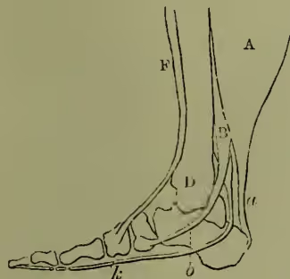
The spot over which the inferior calcaneoscaphoid ligament extends is the weakest in the foot, the astragalus being there unsupported by any bones; additional support is, however, afforded where it is more required by the tendon of a strong muscle, the posterior tibial (fig. 3, B), which passes from the back of the tibia (the chief bone of the leg) round the inner ankle, to be inserted into the lower part of the inner surface of the scaphoid bone. It not unfrequently happens that the astragalus, being either insufficiently supported, or from its being overweighted, descends slightly below its proper level, causing a lowering of the arch and a flattening of the sole of the foot. The defect when slight is known as 'weak ankle;' when more decided it is termed 'flat-foot;' and in extreme cases the bone may descend to such an extent as even to render the inner side of the foot convex when it naturally should be concave.
In the movements of the foot upon the leg we see a striking combination of variety of movement with general security. This combination is effected by the harmonious action of three joints, each of which acts in a direction different from the others. The first of these joints is the ankle-joint, which is formed by the bones of the leg—the tibia and fibula—above, and the astragalus below. At this joint the movements of flexion—i.e. approximation of the toes to the knee, and extension—i.e. pointing the toes to the ground, take place. The second joint is between the astragalus and the heel-bone, and it permits the foot to be rolled inwards or outwards; while the third joint is between the first and second row of tarsal bones—viz. between the astragalus and os calcis behind, and the scaphoid and cuboid bones in front—and allows the degree of curvature of the plantar arch to be increased or diminished within certain limits. The following is the order in which the movements of these three joints occur: the raising of the heel (by the first joint) is accompanied by a rolling of the foot inwards (by the second joint), and by an increased flexure of the plantar arch (by the third joint); and the raising of the toes is accompanied by a rolling of the foot outwards, and a straightening of the sole.
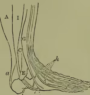
The joints, however, merely allow of movements; they do not effect them: this is the special function of the muscles; and each of the three movements we have indicated is effected by special groups of muscles. The principal of these muscles are shown diagrammatically in figs. 3 and 4, representing the inner and outer sides respectively. The first series of movements is mainly effected by three muscles: viz. (1) muscles of the calf (fig. 3, A), attached above to the bones of the thigh and leg, and below by the Tendo Achilles to the heel-bone; (2) the posterior tibial (fig. 3, B), attached above to the tibia, and below by its tendon to the scaphoid bone; and (3) the short fibular (fig. 4, C), attached above to the fibula, and below by its tendon to the outer metatarsal bone. The calf-muscles, whose tendon is inserted into the heel-bone, are large and very powerful, for in raising the heel they have to raise the weight of the body. The other two muscles, the posterior tibial and the short fibular, turn round the inner and the outer ankle respectively, and are inserted into the inner and the outer edges of the instep; the former being attached to the scaphoid, and the latter to the outer metatarsal bone. They not only assist in raising the ankle, but support it laterally. The muscle whose tendon is on the inner side of the foot (the posterior tibial) effects the two movements which are associated with the raising of the heel-bone—viz. the turning of the foot inwards and the increased flexure of the arch.
The second series of movements—the raising of the toes, the turning of the foot outwards, and the straightening of the sole—are effected by two muscles, the anterior tibial (fig. 3, F) and the third fibular (fig. 4, G), whose tendons pass, one in front of the inner ankle, and the other in front of the outer ankle, to the corresponding edges of the instep, and are inserted into the internal cuneiform and the outer metatarsal bones. These muscles are direct flexors of the tarsus upon the leg; the former raising the inner, and the latter the outer border of the foot.
Another point in the anatomy of the foot that requires notice is the mode of union of the metatarsal with the tarsal bones. In these joints in the fourth and fifth toes a slight revolving motion can take place, which probably enables the outer metatarsals to adapt themselves to inequalities of the ground, and to equalise the distribution of the weight which is thrown upon the foot; while, in the corresponding joints of the three inner toes, scarcely any motion can occur—a provision by which additional strength is given to the inner side of the foot, upon which the weight of the body most directly falls.
The skin of the sole is very tough and strong; and intervening between it and the bones and the strong fascia of the sole of the foot is a thick pad of fat, which acts the part of an air or water cushion in defending the adjacent parts from injurious pressure, and in deadening the jars and shocks that would otherwise be felt in leaping, &c.
A few remarks on the subject of shoes may here be added. The shape of the sole of the natural foot is shown in fig. 5, while the shape after the prolonged use of a badly-made shoe is given in fig. 6. In the foot in its normal state the great toe is seen to be free from the others, and the line of its axis prolonged backwards passes through the centre of the heel; while in the foot distorted by the use of the shoe the line of the great toe is quite altered, and the toes generally—not being able to find room side by side—overlap each other
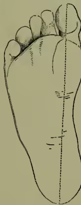
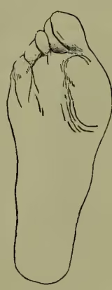
and lose their separate and individual actions; corns, bunions, and ingrowing toe-nails being the natural consequence of this maltreatment. Meyer of Zurich drew attention to the bad treatment which the foot receives from ordinary shoemakers, and pointed out that the great toe should be allowed to have its normal position, and this can be done by making the inner edge of the sole incline inwards, instead of outwards, from the balls of the toes. The accompanying figure (7) gives the outline of a shoe designed under Meyer's superintendence, and shows the difference between it and the usual shape; the latter being indicated by the dotted outline. High heel-pieces tend to make the step less steady and secure, to break down the arch of the foot, to shorten it, and to impair the action of the calf-muscles. A high heel-piece, moreover, places the forepart of the foot at a lower level than the heel; the weight is thus thrown too much in the direction of the toes, and they are thrust forward and cramped against the upper leather of the shoe.
Fig. 7.
Shoe designed by Dr Meyer, the dotted outline being the usual shape.
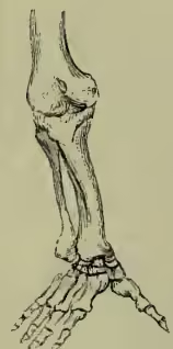
Foot of Gorilla.
If we compare the human foot with the feet of other mammals we find that it presents certain peculiarities, all of which have reference to man's erect posture. The chief peculiarities are (1) the greater relative size of the tarsal bones as compared with the other bones of the foot, and the more perfect formation of the plantar arch, which is higher and stronger than in any of the lower animals. Strength and elasticity are thus combined in the human foot in the highest degree. (2) The great toe is remarkable in man for its size and strength, and for the firm manner in which its metatarsal bone is joined to the other bones so as to render it the main support to the foot. (3) If we compare the human foot with that of the gorilla (fig. 8) or any other Anthropoid Ape (q.v.) we see that the toes are short and small in man in relation to the other parts of the foot, while in the gorilla the toes form the greater part of the foot. Indeed, a reference to fig. 8 shows that the organ in question is rather a hand than a foot, and hence the term quadrumanus as applied to this class of animals. There is scarcely any plantar arch, and the weight of the body bears chiefly on the outer edge of the foot; the digits are long and strong, and the inner one diverges so as to form a thumb rather than a great toe. we see that the large bone in the horse, known as the cannon-bone, which is articulated to the ecto-cuneiform, ce, is the metatarsal of the third toe, to which are articulated the three phalanges of that toe, the last phalanx, 3, being expanded to form the hoof. The small bone popularly known as the splint-bone (not shown in the figure), and articulated to the meso-cuneiform, is the rudimentary or stunted metatarsal of the second toe, 2; and the outer splint-bone, articulated to the cuboid, is the rudimentary metatarsal of the fourth toe, 4; so that in the horse we have
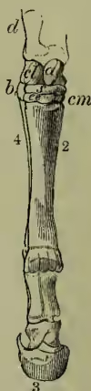
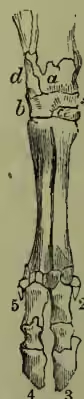
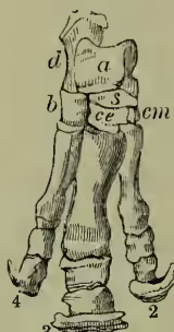
only one toe, the third, sufficiently developed to reach the ground, with mere traces of a second and fourth toe on either side. In the foot of the ox the cuboid, b, is relatively larger than in the horse, and is equal in size to the ecto-cuneiform, ce. The cannon-bone articulates with both these tarsal bones, and hence answers to the metatarsal bones of both the third and fourth digits; it is accordingly found to consist of two distinct bones in the fetus; and in the adult it is divided internally into two cavities, and its original separation is marked out by an external elongated ridge.
Rhinoceros.
Hippopotamus.
Elephant.
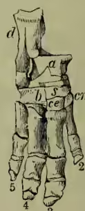
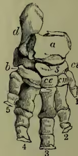
At the lower end are two distinct joints for the phalanges of the third and fourth toes. While in the horse we had the rudiments of the upper parts of two toes (the second and fourth), in the ox we have the rudiments of the lower parts or phalanges of two toes (the second and fifth), forming the 'spurious hoofs,' and marked 2 and 5 in the figure. In the rhinoceros there is one principal toe (the third), as in the horse, with the second and fourth toes in a less developed state; while in the hippopotamus there are two principal toes (the third and fourth), as in the ox, with the second and fifth toes not fully developed. In the elephant there is a fifth digit added, answering to our great toe, and articulating with an ento-cuneiform bone, so that in the foot of this animal we have all the bones occurring in the human foot. Owen concludes from these and similar observations that the course of the simplification of the five-toed foot is, first, a diminution and removal of the innermost toe; next, of the outermost; then, of the second; and lastly, of the fourth; the third or middle toe being the most constant and (in the lower animals) the most important of the five.