Nervous System, the mechanism by which an animal acquires a knowledge of the external world, and by means of which the great functions of the absorption of food, the elimination of waste products from the body, the respiration, circulation, and muscular action, are regulated and controlled. In its simplest form, in some of the lowest animals, it consists merely of nerve-fibres going to and from a small group of nerve-cells; but from this elementary condition there is an ever-increasing degree of complexity till its highest development is reached in man, which alone is included in the following description. In the articles on Birds, Fishes, Reptiles, Mollusca, &c. will be found paragraphs on the nervous systems of the various classes of animals.
The nervous system is composed of a series of organs—nerve-cells, nerve-fibres, and nerve end-organs. The nerve-cells are situated for the most part in the brain and spinal cord, but also in other parts of the nervous system. Their function is either to receive, to send out, or to modify as they transmit, nervous impulses. The nerve-fibres transmit nerve impulses to and from the nerve-cells. For this reason groups of nerve-cells are often conveniently spoken of as nerve centres; their relation to the nerve-fibres being analogous to that of a telegraph-office to the wires connected with it. The end-organs are the special structures for receiving impressions, such as the various organs of special sense, smell, sight, hearing, taste, touch (see NOSE, EYE, EAR, TASTE, TOUCH), and for transforming outgoing impulses into muscular contractions or secretion, &c. The nerve-fibres thus connect either an end-organ with a nerve centre, or two nerve centres with each other. These three sets of organs, nerve centres, fibres, and end-organs, are grouped into two great systems—the cerebro-spinal (fig. 1) and the sympathetic (fig. 10) system. The former is composed of the brain, spinal cord, and the cranial and spinal nerves respectively connected with them. The sympathetic system is formed by a double chain of small swellings, called ganglia, on either side of the front of the spinal column, and connected with each other, with the spinal nerves, and the internal organs by fine nerve-fibres (figs. 7 and 10).
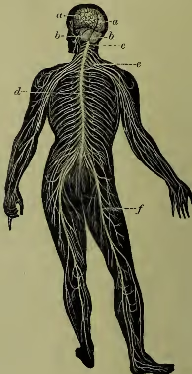
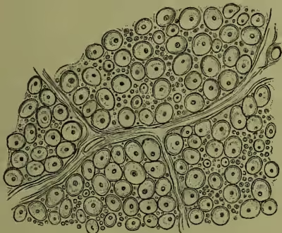
The nerves are whitish cords varying greatly in size. They are composed of nerve-fibres which are bound together by fibrous tissue. This forms a sheath on the outside (the perineurium), and sends processes inwards between the individual nerve-fibres (fig. 4 shows a transverse section of part of a nerve, with the bundles of connective tissue passing inward). In the spinal cord and brain the nerve-fibres are held together by a special kind of connective tissue, called neuroglia.
Structure of Nerve-fibres.—A fibre from a spinal nerve has the following structure. In the centre is a very fine fibre or thread called the axis cylinder. This runs without any interruption along the whole length of the nerve. It can be traced into a nerve-cell at one extremity, and into an end-organ at the other; and there is reason to believe that it is really an outgrowth from a nerve-cell. It is the essential constituent of a nerve, that namely along which the nervous impulse travels. The axis cylinder is in its turn composed of still finer fibrillæ, which may break up into finer nerve-fibres. Except at its origin and termination, the axis cylinder is covered by a tubular membrane called the medullary sheath, or the white substance of Schwann, a whitish substance of a peculiar fatty nature. This is interrupted at intervals of about th of an inch by constrictions which (fig. 2) pass completely through its thickness. When a nerve is stained with nitrate of silver, a black colour is formed at the intersections, and for a short distance along the axis cylinder (see fig. 3, where two are represented). These interruptions are called nodes of Ranvier, after their discoverer, and are supposed to allow of the percolation of lymph to nourish the axis cylinder. When a nerve
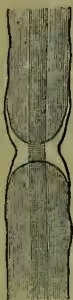
Small part of a nerve-fibre with axis cylinder, surrounded by medullary sheath. The primitive sheath passes over the constriction in the medullary sheath.
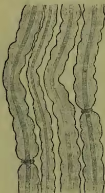
Nerve-fibres, stained with nitrate of silver, showing two nodes of Ranvier.
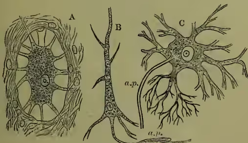
A, from sympathetic ganglion; B, from cerebrum; C, from spinal cord; a.p., axis cylinder process.
is divided transversely, and stained appropriately, the axis cylinder appears as a small point surrounded by a ring of the whitish medullary sheath (fig. 4). This sheath is enclosed by a continuous investment, the primitive sheath. This is colourless, and very delicate, and has a nucleus on its inner side corresponding to each segment of the medullary sheath. In the sympathetic system the medullary sheath is absent; while the fibres of the brain and spinal cord retain the medullary sheath, but want the primitive membrane. The sympathetic fibres are often called gray or non-medullated; the others, white or medullated. The nerve-fibres in the limbs are about th of an inch in diameter; in the brain they may be nearly ten times finer.
The nerve-cells vary greatly in size and in form. Many of them, especially in certain regions of the cortex of the brain, have the shape of an elongated pyramid (fig. 5, B), with fine processes coming off at various points, others are very irregular in outline, but also with numerous processes, one of which can frequently be traced into continuity with a medullated nerve, and hence is called the axis cylinder process, while the others form a fine network before entering another cell or fibre. Such cells are called multipolar (fig. 5, A and C), and are seen best in the anterior horns of the gray matter of the spinal cord. Many cells, again, are bipolar—i.e. they have only two fibres, one at each pole, in connection with them.
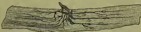
The various end-organs are described under the special sections. Fig. 6 shows the manner in which the fibres of a nerve end in a muscle.
The nerves arising from the brain are arranged in twelve pairs. The first, or olfactory, is the nerve of smell. The second, or optic, is the nerve of sight. It arises from the retina, meets with its fellow in the optic chiasma, and is distributed half to each side of the brain, terminating partly in the corpora quadrigemina (for the reflex movements of the eye), and partly in the optic thalamus, passing thence to the occipital lobe of the cerebrum (for the sense of sight). The third or oculo-motor nerve arises under the corpora quadrigemina, and passes to all the muscles of the eye except two, which are supplied by the fourth and sixth pairs. The fourth nerve, arising immediately behind the third nerve, supplies the superior oblique muscle of the eye; while the sixth pair, arising from a nucleus near the middle of the floor of the fourth ventricle, supplies the external rectus muscle of the eye. The fifth pair has a very long origin from a point at the level of the third nerve down to the upper part of the spinal cord. It is the motor nerve to the muscles of mastication, and the sensory nerve to the face, front of the head, teeth, tongue, and is the nerve of taste of the anterior part of the tongue. It is this nerve which is concerned in neuralgia of the head and face and teeth. The seventh pair arises from the lower part of the pons Varolii (see BRAIN), and is the motor nerve to the facial muscles of expression. Injury to or disease of this nerve causes facial palsy, or Bell's paralysis. The eighth pair, or auditory nerve, supplies the internal ear. It is divided into two parts, one of which supplies the cochlea, and is the nerve of hearing proper, while the other supplies the semicircular canals, and is concerned in the maintenance of the equilibrium of the body. The nerve arises from the lateral and posterior part of the pons Varolii and medulla oblongata. The ninth pair, or glosso-pharyngeal nerve, is the special nerve of taste, and supplies the hinder third of the tongue, with the taste bulbs of which it is connected. The tenth pair, or pneumogastric nerve, has a very wide area of distribution to the lungs, heart, stomach, &c.; it is partly motor and partly sensory in function. The eleventh pair, or spinal accessory nerve, is the motor nerve to the larynx, and to certain muscles in the upper part of the neck. These three nerves arise from a groove in the side of the medulla oblongata and upper part of the spinal cord. The twelfth pair, or hypoglossal nerve, is the motor nerve of the tongue. Its origin is near the floor of the fourth ventricle, close to the middle line, and it emerges from the anterior surface of the medulla oblongata in a shallow groove between the anterior pyramids and the inferior olivary body (see BRAIN).
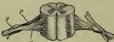
The spinal nerves arise from the spinal cord in pairs, thirty-one in number, and are named according to their relation to the vertebrae—cervical, dorsal, lumbar, and sacral. Their mode of origin will be understood from fig. 7, which represents diagrammatically the first part of their course, and on one side their relations with the sympathetic system—C 1-8 represents the eight pairs of cervical nerves; D 1-12, the twelve dorsal pairs; L 1-5, the five lumbar pairs; and S 1-6, the six sacral pairs of nerves. Each spinal nerve arises by two roots, an anterior and a posterior (fig. 8, a and p; see also SPINAL CORD). These roots pass outwards, and unite before they leave the spinal canal. Before their union a small oval swelling is found on the posterior root, and is called its ganglion, g. The united nerve leaves the spinal canal by a small aperture between adjacent vertebrae. It almost immediately gives off a fine medullated nerve to its corresponding sympathetic ganglion, a branch which can be traced into one of the internal organs. It also receives from the ganglion a non-medullated or gray fibre, which is distributed to the muscular coat of the blood-vessels, especially the arteries. The nerve thus altered passes outwards, dividing as it goes to send its ultimate branches into the fibres of the muscles, into the cells of the skin and connective tissues, tendons, and bones. In the dorsal region each nerve passes to its distribution without entering into connection with its neighbours, but in the cervical, lumbar,
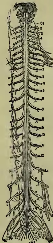
and sacral regions the nerves split up and form new junctions with each other, or plexuses as they are called. (These are indicated in fig. 1, and on the right-hand side of fig. 7, but the detailed description of them is impossible within the limits of this article.)
Functions of the Spinal Nerves.—Sir Charles Bell discovered that division of the anterior roots was followed by loss of power of voluntary motion, and that division of the posterior roots destroyed the power of sensation. He termed the anterior root motor, and the posterior sensory. It has since been ascertained that the anterior roots carry outwards other impulses that do not result in motion, and that the posterior roots carry inwards impulses which may not result in sensation. Therefore, it is more correct to term these roots respectively afferent and efferent. If the anterior root be divided between the point of its origin from the cells of the anterior horn of the spinal cord and its junction with the posterior root, the part unconnected with the cord will waste along the whole length of the nerve, and the muscles which it supplies will waste also. The cells in connection with the anterior roots, therefore, not only send out motor impulses, but exert a nutritive or trophic influence on the nerve and muscle. Division of the posterior root beyond its ganglion is followed by wasting of the corresponding fibres of the nerve to their ultimate termination. If the root be cut between the ganglion and the spinal cord, the part attached to the ganglion remains unaltered, while that connected with the spinal cord wastes. This shows that the ganglion of the posterior root exerts a trophic influence on the fibres connected with it. If the nerve be divided after the junction of the two roots, the whole of the nerve farthest from the spinal cord will waste.
The afferent nerve impulses which pass along the posterior roots comprise those which give rise to the sense of touch, pain, and temperature, and to reflex movements of various kinds without necessarily exciting our consciousness, such as those concerned with the maintenance of the equilibrium of the body, and with the functions of the internal organs.
Reflex Action.—By this we mean an action brought about directly by the influence of an afferent impulse quite independently of voluntary control. For such an action four elements are necessary: (1) afferent fibres, (2) nerve cells or centres, (3) efferent fibres, (4) muscle fibres. The impulse travels up the afferent fibres and stimulates the nerve-cells to send an impulse along the efferent fibre to the muscles. If any of these four factors is absent, the reflex action cannot take place. A familiar example is the moving of the foot as the result of tickling the sole. The afferent impulse passes up the nerves to the nerve centres in the spinal cord, which send outwards direct to the muscles motor impulses, which often cannot be controlled by the will.
Automatic Action.—When movement is brought about by an impulse originated in a nerve centre itself, without the influence of an afferent stimulus, it is called automatic or spontaneous. Such actions are apt to occur rhythmically, such as the action of the heart.
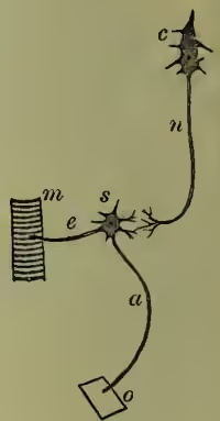
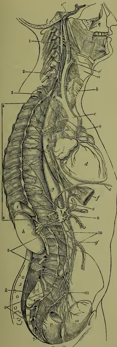
Voluntary Actions.—In these the outgoing impulses originate in the nerve-cells in the motor area of the brain, and pass down the opposite side of the spinal cord to the nerve-cells in its anterior horn. From thence they are transmitted by the efferent nerves to the muscles. Fig. 9 will explain the relation of voluntary to reflex action: c is a nerve-cell in the brain; n, the nerve-fibre in the spinal cord which transmits the nerve impulse originating in c to s, a cell in the anterior horn of the spinal cord which forwards it through a nerve, e, to a muscle, m. The reflex arc is represented by o, an end-organ; a, an afferent nerve; s, a nerve-cell; e, an efferent fibre; m, a muscle. If c or n, or both, be destroyed by disease, the power of voluntary motion will be lost, but the reflex arc, o, a, s, e, m, remains intact. We can sometimes control or 1, 2, 3, the superior, middle, and inferior cervical ganglia; 4, the two lines from this figure include the twelve dorsal ganglia; 5, include the four lumbar ganglia; 6, include the five sacral ganglia; 7, the ganglion impar; 8, cardiac plexus; 9, solar plexus; 10, aortic plexus; 11, hypogastric plexus; a, the larynx; b, the trachea; c, arch of the aorta; c', external carotid; c'', internal carotid; d, the heart; e, e', the diaphragm; f, the cardiac end of the oesophagus; g, thoracic, and g', abdominal aorta; h, the kidney; i, the supra-renal capsule; k, the sacrum; l, the section of base of the skull; m, the bladder; n, the lower portion of the rectum. repress a reflex action voluntarily. The cell c in the brain can so act on the cell s as to hinder or inhibit its ordinary response to a stimulus, and when the cell in the brain is destroyed the cell s is more easily stimulated reflexly, apparently because some restraining influence is removed. Such a restraining action is exerted on the heart by the pneumogastric nerve, which, when stimulated, slows or stops its movements; and in the same way the secretion of glands may be interfered with by abnormal stimulation of their nerve centres. This influence is termed inhibitory action, and is one of great importance.
Secretory Nerves.—The nerves which induce the various acts of secretion leave by the motor cranial nerves, or by the anterior roots of the spinal nerves; but the description of their distribution, &c. must be relegated to the article SECRETION.
The sympathetic system is, as already stated, composed of a series of ganglia situated on either side of the spinal column along its whole length. In the dorsal, lumbar, and sacral regions the ganglia correspond in number to the vertebrae; but in the cervical region there are only three, of such large size, however, that they are generally supposed to represent the fusion of a number of ganglia. Below, the two chains unite in front of the coccyx in a single ganglion. These ganglia are formed of multipolar nerve-cells (fig. 5, A), and are united with each other by gray nerve-fibres. Each ganglion gives to its corresponding spinal (or cranial) nerve a gray non-medullated nerve, and receives from it a fine white medullated nerve. The fibres of distribution may be studied in fig. 10. They pass to the blood-vessels and to the mucous membranes and muscular coats of the various internal viscera, and become united with each other in fine networks or plexuses, on many of which nerve-cells or ganglia are situated.
The sympathetic chain is continued upwards as a fine plexus of nerves on the internal carotid artery, on the various branches of which it is distributed. From the superior cervical ganglion also fibres pass to the various arteries in the neck and face, and to form, along with the pneumogastric and glossopharyngeal nerves, the pharyngeal plexus on the muscles and mucous membrane of the pharynx.
From some of the cervical and upper thoracic ganglia fibres pass into the chest, to form also, along with the pneumogastric nerve, two important plexuses, named pulmonary and cardiac, from which branches pass to the lungs and heart, and undoubtedly influence their functions. From the thoracic ganglia also arise the three splanchnic nerves which pass into the abdomen to enter the solar or epigastric and the renal plexus. The solar plexus is situated at the pit of the stomach, and is connected with two large semilunar ganglia, which send branches to all the blood-vessels and to all the organs within the abdomen. It is owing to the relations and functions of the solar plexus that blows in this region are so dangerous. The hypogastric plexus arises from the lumbar ganglia, and sends branches to the blood-vessels and to the organs in the lower part of the abdominal cavity, more especially the organs of generation, the lower bowel, and the bladder.
The functions of the sympathetic system are still imperfectly understood. It supplies fibres to the muscles of blood-vessels to regulate their calibre; hence these fibres are called vaso-motor. The vaso-motor centre is situated, not in the sympathetic system, but in the medulla oblongata. The path for the fibres passes down the spinal cord, which it leaves by the anterior roots at various levels to pass along the white communicating branches into the sympathetic ganglia. From these ganglia the vaso-motor nerves for the internal organs pass into the various plexuses just described, while those for the vessels of the limbs and trunk return to the spinal nerve by the gray communicating nerve. Further, the muscular movements and secretions of all the internal organs are regulated through the sympathetic system.
Of the nature of nerve energy we know little. The nervous impulse travels along a nerve in man at a rate of about 34 yards per second, and it is accompanied by certain electrical changes in the nerve, but nerve energy is not identical with electrical energy. It is probably accompanied by molecular changes in the structure of the nerve as yet inaccessible to our means of investigation. In like manner the origination or discharge of nerve impulse in a cell is probably induced by similar but more active changes in its substance.
See Quain's Anatomy, Foster's Physiology, Landois and Stirling's Physiology, and Obersteiner's Anatomy of the Central Nervous System (translated by Hill).