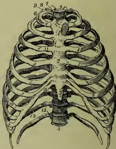
Ribs are elastic arches of bone, which, with the vertebral column behind, and the sternum or breast-bone in front, constitute the osseous part of the walls of the chest. In man there are twelve ribs on each side. The first seven are more directly connected through intervening cartilages with the sternum than the remainder, and hence they are termed vertebro-sternal or true ribs; while the other five are known as false ribs, and the last two of these, from being quite free at their anterior extremities, are termed floating ribs. A glance at a skeleton, or at a plate representing the articulated 1 and 2 are the upper and the middle parts of the sternum or breast-bone; 3, its ensiform cartilage; 4, the first dorsal, and 5 the last (or twelfth) dorsal vertebra; 6, the first rib; 7, its head; 8, its neck, resting against the transverse process of the first dorsal vertebra; 9, its tubercle; 10, the seventh or last true rib; 11, the costal cartilages of the true ribs; 12, the last two false ribs or floating ribs. bones, will show that the ribs vary very considerably both in their direction and size. The upper ribs are nearly horizontal, but the others lie with the anterior extremity lower than the posterior; this obliquity increasing to the ninth rib, and then slightly decreasing. They increase in length from the first to the eighth, and then again diminish. The spaces between the ribs are termed the intercostal spaces. On examining a rib taken from about the middle of the series we find that it presents two extremities (a posterior or vertebral, and an anterior or sternal), and an intervening portion, termed the body or shaft. The posterior extremity presents a head, a neck, and a tuberosity. The head is marked by two concave articular surfaces divided by a ridge, the lower facet being the larger. These surfaces fit into the cavity formed by the junction of two contiguous dorsal vertebrae, and the ridge serves for the attachment of a ligament. The neck is a flattened portion proceeding from the head; it is about an inch long, and terminates at an eminence termed the tuberosity or tubercle, from whence the shaft commences. On the lower and inner part of this tubercle is a small oval surface, which articulates (as shown in fig. 2) with a corresponding surface on the upper part of the transverse process of the lower of the two vertebrae with which the head is connected. The shaft presents an external convex and an internal concave surface. A little external to the tubercle the rib is bent to form the angle, from which point the rib passes forwards and outwards, ultimately curving inwards to join its costal cartilage. The upper border of the rib is thick and rounded, while the lower border is marked by a deep groove, which lodges the intercostal vessels and nerve.
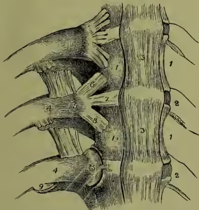
The ribs of Mammals are mostly connected, as in man, with the bodies of two vertebrae, and with the transverse processes of the posterior one. In the Monotremata, however, they articulate with the vertebral bodies only; while in the Cetacea the posterior ribs hang down from the transverse processes alone. Their number on each side corresponds with that of the dorsal vertebrae. The greatest number, twenty-three, occurs in the two-toed sloth, while in the Chiroptera eleven is the ordinary number. In Birds each rib articulates by means of a small head with the body of a single vertebra near its anterior border, and with the corresponding transverse process by means of the tubercle. Moreover, each rib possesses a 'diverging appendage,' which projects backwards over the next rib, so as to increase the consolidation of the 1, 1, dorsal vertebrae; 2, 2, intervertebral cartilages; 3, 3, anterior common ligament; 4, neck, and 5 head of rib; 6, 7, 8, flat bundles of ligamentous fibres (removed in the lowest rib, shown in the figure); 9, articulation between the tubercle of the ribs and the transverse vertebral process. thoracic framework, necessary for flying. The dorsal vertebrae here never exceed eleven, and are commonly seven or eight in number, and the ribs proceeding from them are connected with the sternum not by cartilage, as in mammals, but by true osseous sternal ribs, which are regularly articulated at one end with the sternum, and at the other with the termination of the spinal ribs. In the Chelonian Reptiles the ribs (as well as the vertebra and the sternum) deviate remarkably from the normal type, the lateral parts of the carapace consisting mainly of anchylosed ribs united by dermal plates. In the Crocodiles there are only twelve pair of true or dorsal ribs; while in the other Saurians, and in the Ophidians, the ribs are usually very numerous. In the Frogs there are no true ribs, the reason probably being that any bony element in their thoracic walls would interfere with the enormous thoraco-abdominal enlargement which these animals periodically undergo at the breeding period.
In the language of the comparative anatomist, a rib is to be regarded as a Pleurapophysis—one of the elements of a typical Vertebra (q.v.).
Fracture of the Ribs is a very common surgical accident, resulting from blows or falls upon the chest. Ribs may, moreover, be broken by mere pressure, as when persons are severely crushed in a crowd; and instances are on record in which, in the case of aged persons, the ribs have been actually fractured in violent coughing. The treatment consists in the application of a broad flannel roller round the chest, so tightly as to prevent, as far as possible, all movement of the ribs, and to render the respiration abdominal rather than thoracic. The bandage must be prevented from falling by the addition of shoulder-straps; and in order to prevent the shoulder-blade from moving, and thus disturbing the broken ribs, some surgeons confine the arms to the side of the body. If one or both of the extremities of the fractured rib should perforate both layers of the pleura and wound the lung, or in rare cases when only the parietal layer of the pleura is injured, if the skin is also perforated, air may escape in the act of inspiration from the lung or from the exterior into the pleural cavity, and thence through the wound in the costal pleura into the cellular or areolar tissue of the trunk, giving rise to Emphysema (q.v.), in the form of a soft puffy swelling that crepitates and yields on pressure.