Skeleton, a general term for the more or less hard parts of animals, whether forming an internal supporting framework—an endoskeleton, or an external exoskeleton, often useful as armour. The term includes so many different kinds of structure and material that it is necessary to take a survey of representative types.
Skeleton of Invertebrates.—Many of the Protozoa have shells of lime (see FORAMINIFERA), or of flint (see RADIOLARIA), or of some organic substance, such as acanthin. These are formed by the living matter of the units, in the case of the lime and flint shells from materials absorbed from the surrounding water, but in what precise way we do not know. Almost all Sponges (q.v.) are supported by loose or firmly fused spicules of lime or of flint, or have, as in the bath-sponge, an interwoven supporting skeleton of 'horny' fibres. The spicules or fibres are formed by cells in the middle stratum of the sponge. Among Cœlenterates various forms of skeleton, both external and internal, both limy and 'horny,' are represented by the different kinds of Corals (q.v.). With few exceptions these skeletons are produced by cells belonging to the outer layer or ectoderm of the animal. Worms have little that can be called a skeleton, although some authorities would compare the sheath of the proboscis in Nemertea (q.v.) to the notochord of Vertebrates. The tubes, calcareous or otherwise, in which many sedentary worms are sheltered, have no vital connection with the animals which make and inhabit them. Echinoderms tend to be very calcareous; lime is deposited in the mesodermic tissue of the body in almost any part, though predominantly near the surface. Most Arthropods have well-developed exoskeletons, cuticles formed from the epidermis, consisting in great part of an organic basis of chitin, on which, in Crustaceans and most Myriopods, carbonate of lime is also deposited. As this cuticle is not always restricted to the outside of the animal, but sometimes extends inwards, an apparent endoskeleton arises—e.g. in the lobster, the king-crab, and the scorpion. Most Molluscs have shells in which carbonate of lime occurs along with an organic basis conchiolin, and in cattle-fish there is a remarkable development of cartilage around the nerve-centres in the head—an analogue of the skull in Vertebrate animals. From this rapid survey it will be seen that the skeletons of Invertebrates are very varied alike in structure and in composition; if we except a few doubtful hints of a supporting axis, there are no homologies between the skeletons of Invertebrates and Vertebrates; to the latter, moreover, that form of tissue which we call bone is exclusively restricted.
Skeleton of Vertebrates.—Here we must distinguish first of all between the external exoskeleton and the internal endoskeleton. The scales of fishes, the scales and scutes of reptiles, the scales, claws, and even feathers of birds, the remarkable bony armature of armadillos, the scales of pangolins, the claws of carnivores, the quills of porcupines, and even the hair of ordinary mammals illustrate the variety of structures which may be included within the anatomical conception of an exoskeleton. All these structures are formed in the epidermis, or in the dermis, or in both combined. Tortoise-shell and the scales of reptiles are epidermic; the scutes of crocodiles and the plates covering armadillos are dermic; the scales of Elasmobranch and Ganoid fishes are due to both layers. But it is difficult to carry out any rigidly logical classification. Thus, the dorsal shield of a tortoise is physiologically an exoskeleton, but structurally it is in great part formed from the dorsal vertebrae and from what in other animals form the ribs. The ventral shield of a tortoise is formed from dermal bones, and the so-called abdominal ribs of crocodiles arise as ossifications in the fibrous tissue which lies underneath the skin and above the muscles. The teeth of Elasmobranch fishes are undoubtedly homologous with the dermal denticles or skin-teeth which occur over the skin, and the teeth of mammals are started by enamel germs which sink in from the epidermis of the mouth.
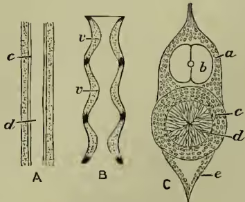
The Vertebral Column.—In a primitive Vertebrate animal like the lancelet the body is supported by a median dorsal axis, and, apart from slight supports for the mouth, the pharynx, and the median fin, this is all the skeleton. The median dorsal axis, which in the lancelet has not even the firmness of cartilage, is called the notochord, and is one of the constant characteristics of the skeleton of Vertebrates. There is a hint of it in the proboscis of Balanoglossus (q.v.) and in Cephalodiscus (q.v.); it is more distinct in the tail of young Ascidians (q.v.), and persists throughout life in Appendicularia; in the lancelet, in the hag, and in the young lamprey it is an unsegmented rod with a yet no vertebral bodies. These begin in the Elasmobranch fishes, in which the notochord is, in part at least, constricted by the encroachment of its sheath, and divided into vertebrae. In the bony Ganoids, such as Lepidosteus, the vertebrae are ossified, and so they are more or less thoroughly in all the higher Vertebrates. Yet it is not the notochord which is ossified, but its sheath; the notochord in all higher Vertebrates being merely a provisional structure, an internal scaffolding around which its 'substitute,' the backbone, is built. Each vertebra generally consists of the substantial body or centrum, the neural arches which form a tube for the spinal cord and meet above it in a neural spine, the transverse processes which project laterally and are usually connected with ribs, and the articular processes which bind vertebra to vertebra so that a firm and yet flexible backbone results.
A breastbone or sternum to which the ribs are ventrally united occurs in many Reptiles and in all Birds and Mammals. It arises from a cartilaginous tract uniting the ventral ends of the ribs. In Amphibians also there is a breastbone, but there are no distinct ribs, and it is not certain that we can compare this sternum with that of higher Vertebrates.
The Skull.—In a young Vertebrate animal the cavity in which the brain lies is surrounded by a membranous sheath, but this is gradually replaced, first by a gristly brain-box, and afterwards in most cases by an almost entirely bony skull. Let us first consider the gristly brain-box or chondro-cranium. (a) Its foundation is formed from two pairs of cartilaginous plates—posterior parachordals and anterior trabeculae—which lie beside and in front of the notochord. These are extended upwards by a further formation of cartilage; the end of the notochord may also help a little: the result is a cartilaginous brain-box. (b) But to the sides of this are added a pair of cartilaginous nasal capsules in front, and a similar pair of auditory capsules behind. (c) About the mouth there are some lip or labial cartilages, which may help in forming the skull; but much more important is a series of cartilaginous 'branchial arches' (never more than eight pairs), which loop round the pharynx, running between the primitive gill-clefts. Of these arches the two most anterior, which are called the mandibular and the hyoid arches, are of great importance in the development of the skull; the others form supports for the pharynx, and are permanently important only in Fishes and in gilled Amphibians. In Elasmobranch fishes the mandibular and hyoid arches do not form any direct part of the gristly brain-box, but in the Teleosts and thence onwards they, or the bones which replace them, contribute directly to the upbuilding of the skull. To follow the history of the arches, which undergo numerous transformations, is one of the most difficult and interesting tasks of comparative anatomy. (d) When a bone develops in direct relation to a pre-existent cartilage which it replaces, it is often called a primary or 'cartilage bone'; and there are many regions of the cartilaginous brain-box which in the course of development are thus replaced by bones. But there are other bones which develop independently of pre-existent cartilage. They invest the cartilaginous brain-box on its roof, on its floor, and on its sides. They are comparable to the dermal ossifications or scutes which occur in Ganoid fishes and many other animals, and they are often called secondary or 'membrane' bones. In structure they are of course indistinguishable from 'cartilage bones'; in origin too they are in one way the same, for all bones arise from a (periosteal) membrane of bone-making cells; but 'cartilage bones' are at one time represented by cartilages, whereas 'membrane bones' never are. eight pairs), which loop round the pharynx, running between the primitive gill-clefts. Of these arches the two most anterior, which are called the mandibular and the hyoid arches, are of great importance in the development of the skull; the others form supports for the pharynx, and are permanently important only in Fishes and in gilled Amphibians. In Elasmobranch fishes the mandibular and hyoid arches do not form any direct part
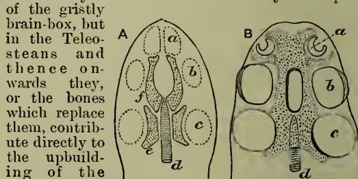
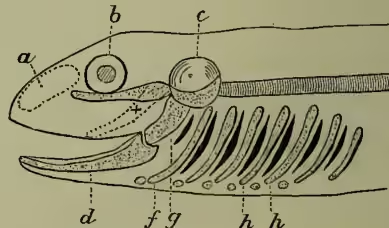
To sum up, the skull is formed (a) from the parachordals and trabeculae at the end of the notochord, (b) from the adjacent sense-capsules of the nose d and g, parts of first or mandibular arch; f, part of second or hyoid arch; behind these the arches (h) separating gill-clefts; a, b, c, the nasal, optic, and auditory capsules. and the ear, (c) from the more or less intimately associated mandibular and hyoid arches, (d) from the ossification of the gristly brain-box due to (a), (b), and (c), but also from 'membrane bones' or existing bones which arise independently of pre-existent cartilages. There is no skull in Tunicates or in the lancelet; it is cartilaginous in Cyclostomata and Elasmobranchs; centres of ossification and investing bones begin with the Ganoid fishes, and are numerous in Teleosteans and in all higher animals. In the development of the individual there is a parallel progress.
Theory of the Skull.—About the beginning of the 19th century Oken and Goethe independently suggested what is known as the vertebral theory of the skull—an undoubtedly suggestive theory, to which Owen lent the strength of his authority, but which has been disproved by the subsequent discoveries of comparative anatomy and embryology (see SKULL).
The Appendicular Skeleton.—Somewhat apart from the axial skeleton are the limbs and the girdles to which these are attached. No secure conclusion has yet been reached as to origin of the limbs of Vertebrates. In the simplest forms—the Tunicates, the lancelet, the Cyclostomata—there are none, and there is a very marked difference between the fin-like limbs of fishes and the fingered and toed limbs which occur in almost all higher backboned animals. According to Gegenbaur, the pectoral and pelvic girdles are structures comparable to the branchial arches, and he supposes that the primitive limbs were made up of modified fin-rays comparable to those which support the unpaired fins of fishes. According to Dohrn the limbs are residues of a longitudinal series of segmentally arranged outgrowths, perhaps comparable to the appendages of a typical Annelid worm (see FISHES, VERTEBRATA).
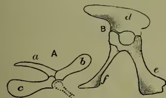
The pectoral or shoulder-girdle consists of a dorsal shoulder-blade or scapula, a ventral coracoid, with the articulation for the arm between them, and of a forward-growing collar-bone or clavicle. The pelvic or hip-girdle consists on each side of a dorsal ilium, a ventral ischium, with the articulation for the leg between them, and of a third pubic portion. The fore-limb—from Amphibians onwards—consists of a humerus articulating with the girdle, a lower arm composed of radius and ulna lying side by side, a wrist or carpus of several elements, a hand with metacarpal bones in the palm and with fingers composed of several joints or phalanges. The hind-limb—from Amphibians onwards—consists of a femur articulating with the girdle, a lower leg composed of tibia and fibula lying side by side, an ankle region or tarsus of several elements, a foot with metatarsal bones in the sole and with toes composed of several joints or phalanges. Distinct from all the other bones are a few little 'sesamoids' which are occasionally developed within tendons and near joints, notably, for instance, the knee-pan or patella. skeleton (fig. 5). Altogether there are more than 200 bones, but some which are originally distinct become fused with their neighbours.
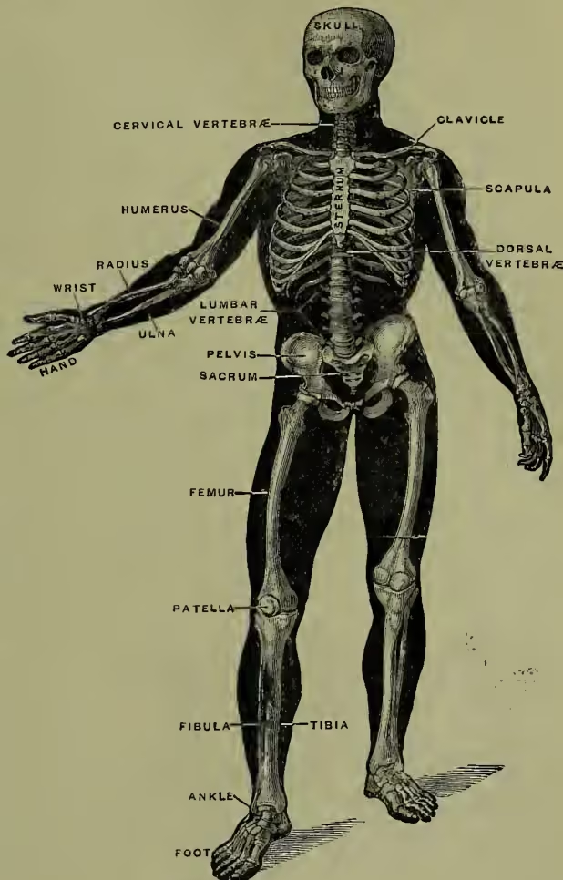
In the vertebral column there are originally thirty-three vertebrae, but in adult life the normal number is twenty-six, for, while the first twenty-four remain distinct, five (the twenty-fifth to the twenty-ninth inclusive) unite to form the sacrum supporting the hip-girdle, and the four hindmost fuse more or less completely in a terminal tail-piece or coccyx. Seven cervicals support the neck; twelve dorsals form the greater part of the back and bear ribs; five lumbars occur in the loins; these are followed by the sacrum and the coccyx (see SPINAL COLUMN).
The ribs, or elastic arches of bone which bound the chest, are normally twelve on each side. Most of them articulate dorsally with the bodies of two adjacent vertebrae and with the transverse processes of the posterior one; ventrally the first seven pairs are connected with the median breastbone by means of intervening cartilages, while the posterior five pairs are more or less free (see RIBS).
The skull consists in early adult life of twenty-two separate bones, but originally there were more, and as life continues the number may be further reduced by fusion. For the various bones, see SKULL.
The skeleton of the arm includes thirty bones—in the upper arm the humerus, which articulates with the shoulder-girdle; in the forearm the radius and ulna, which articulates with the humerus at the elbow; the wrist of eight carpal bones; the five metacarpals of the palm; the five digits, of which the four fingers have each three joints or phalanges, while the thumb has two. The important bone of the pectoral girdle is the shoulder-blade or scapula. To this, at the shoulder-joint, there is fused a small beak-like bone—the coracoid—which is separate in Birds and Reptiles, but reduced to a mere process of the scapula in all Mammals except the Monotremes. Stretching from the breastbone to shoulder-blade is the curved collar-bone or clavicle.
The skeleton of the leg also includes thirty bones—in the thigh the femur, which articulates with the hip-girdle; in the lower leg the shin-bone or tibia and the splint-bone or fibula, which articulate with the femur at the knee-joint, where there lies a little ‘sesamoid’ bone—the patella; in the ankle region seven bones, then five metatarsal bones forming the sole of the foot, and five toes with the same number of phalanges as in the fingers. The pelvic girdle consists in early life of three paired bones—large dorsal ilium, a posterior ischium, an anterior pubis on each side—but these unite about the twenty-fifth year into single haunch-bone, with the socket of which the thigh articulates. See ARM, HAND, SHOULDER, FOOT, LEG, PELVIS, RIBS, SKULL, and SPINAL COLUMN; for literature, see the works referred to in the article ANATOMY. Skeletons other than human will be seen under the headings of ANTHROPOID APES, BAT, BIRD, ELK, FISHES, HESPERORNIS, ICHTHYOSAURUS, IGUANODON, MEGATHERIUM, PLESIOSAURUS, PTERODACTYL, &c.; and skulls at BABIROUSSA, CARNIVORA, DOG, RODENT, &c.