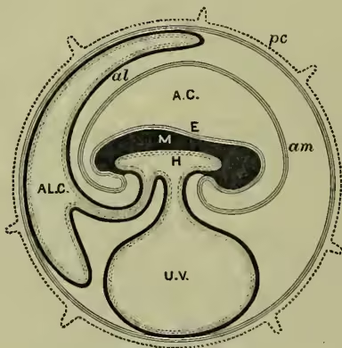
Allantois, a sac-like foetal membrane formed in the embryos of reptiles, birds, and mammals, as an outgrowth from the posterior end of the alimentary tract, just in front of the anus. While it never really occurs in animals lower than reptiles, it may be fairly said to be represented in the fish Lepidosiren and in the amphibia by a urinary bladder. In all higher animals, however, it is precociously developed and modified for other purposes. It forms a large sac, enveloping the embryo outside the Amnion (q.v.). In reptiles and birds the surface is traversed by an abundant network of blood-vessels, and the important function of embryonic respiration is thus discharged. In placental mammals a further functional change (see FUNCTION) is exhibited, since the allantois aids in the development of the chorion, and this again is intimately connected with the walls of the uterus, in the structure known as a Placenta (q.v.). The blood of the embryo is exchanged with the blood of the mother, and embryonic nutrition is thus effected. The precocious urinary bladder is thus respiratory in reptiles and birds, and nutritive in mammals. In amphibia, the allantois persists as stated; in reptiles, the stalk dilates to form the permanent bladder; in birds, the whole atrophies. In mammals, the stalk of the allantois (1) dilates to form the bladder, while the lower portion (2) narrows to form the urethra. The urachus, or stalk connecting the bladder to the ventral wall of the body, is also of allantoic origin.