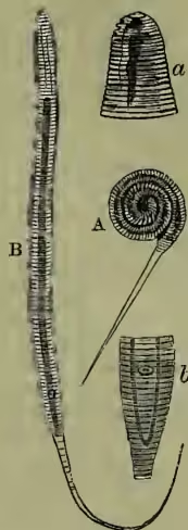
Guinea-worm, known also as Filaria Medinensis, or F. Dracunculus, is a parasitic animal that seems to have been known from the earliest times. Plutarch quotes a passage from a still earlier author which seems clearly to refer to this worm. But our knowledge of its natural history is still very deficient, and we are at present only acquainted with the female. The body of this animal is slender, cylindrical, and somewhat compressed, and is of the thickness of pack-thread, except at the posterior extremity, where it is somewhat attenuated. It is opaque, of a milk-white colour; on each side there is a longitudinal line; and when examined by the microscope it is seen to be marked with numerous transverse striae. The anterior extremity is obtuse and the mouth circular and beset with four acute spines. The length of the worm varies from less than half a foot to three yards. On examining an adult specimen, extracted by Malgaigne in Paris in 1854, Robin found no trace of intestine, or of any organ except a very thin sheath (a uterus or oviduct), which was filled with young animals rolled up in coils, with the tail occasionally projecting outwards (see A in the figure). In these young animals we can trace the course of the intestinal canal, which apparently becomes subsequently obliterated by the excessive development of the generative organs and the eggs.
This worm is indigenous only in certain hot countries, and its geographical distribution is regulated by laws into which we have no insight, save that heat and moisture are necessary for its production. Among places as especially notorious for its occurrence are Senegal, Gaboon, the banks of the Ganges, Bombay, the peninsula of India, Persia, Arabia Petraea, the south coast of the Red Sea, the region round the Caspian Sea, Upper Egypt, Abyssinia, certain districts of Nubia, the swampy regions of the White Nile, and Guinea. It has been introduced into certain parts of America by negro slaves. The disorder occasioned by these worms frequently becomes an epidemic in years of heavy rain, and especially in marshy districts. It appears also to be connected with the season, being especially prevalent in the East Indies during the rainy season, and in Upper Egypt shortly after the regular inundation of the Nile.
The mode of production of this parasite in the human body is not known with certainty. The probability is that the young animals while still very minute penetrate the skin, although by what mechanism they can effect their lodgment we do not know. Having gained an entrance into the body, the guinea-worm takes a considerable time to develop. This period varies from two months to a year or even two years. The presence of the worm often produces no annoyance for a considerable time after it has been detected; at other times it gives rise to emaciation, and possibly even death from exhaustion. As a general rule the vesicles caused by the inflammation excited by the presence of the worm open spontaneously in a few days, and two or three inches of the anterior end of the animal come forth. This end is gently pulled, and coiled round a little roll of linen or a small stick, and this is fastened over the wound with sticking-plaster and a compress. The extraction is repeated twice a day by rotating the substance round which the worm is twisted, and the operation is often not completed in less than two, three, or more months. From the most ancient times the tearing of the worm has been regarded as a very dangerous accident. It undoubtedly gives rise to violent swelling, fever, sleeplessness; and shortening and deformities of the legs, lingering fistula, mortification, and death (sometimes even sudden death) must be reckoned amongst the notable consequences of breaking the worm. See Quain's Dictionary of Medicine;
Linn. Trans. (1863); also the writings of Owen, Cobbold, and Bastian.