Heart, the central organ of the circulatory system, acting as a force and suction pump in relation to the blood-vessels. It always lies dorsally in Invertebrates, ventrally in Vertebrates, and arises from the strong development of one or more blood-vessels. In Vertebrates, the resulting cylinder, lying in the throat region of the embryo, is divided into receiving and expelling portions, auricle and ventricle respectively, and the whole is enclosed in a more or less marked cavity or ensheathing double bag, the pericardium. By curvature and folding, by formation of partitions and ingrowth of valves, the three or four chambered hearts of the higher vertebrates arise. It will be enough to describe the general structure and function of the heart in man.
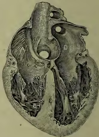
The human heart lies ventrally in the chest, between the two lungs; it has a broad end or 'base' directed upwards and backwards, and a pointed end or 'apex,' turned downwards, forwards, and to the left; it is kept in position by the attachment of the ensheathing pericardium to the upper surface of the Diaphragm (q.v.), and by the large blood-vessels which enter or leave its four chambers; its total size is approximately equal to that of its owner's closed fist. There are two receiving chambers or auricles, of which the right receives all the impure blood brought by the vena cava from head and body and by the coronary vein from the substance of the heart itself, while the left is filled with purified blood brought by the pulmonary veins from the lungs. The auricles pass their contents to the two driving chambers or ventricles, of which the right pumps the impure blood to the lungs, and the left sends the pure blood to the head and body. The ventricles are larger than the auricles, and have strong muscular walls proportionate to their harder work. The left ventricle is stronger than, and partially surrounded by, the thinner right chamber. The right auricle opens into the right ventricle by an aperture guarded by a triple (tricuspid) valve, whose three membranous lappets are attached to tendinous cords (chordae tendineae) arising from muscular processes (musculi papillares) on the walls of the ventricle. The opening from the left auricle into the left ventricle is similarly guarded by a double (mitral or bicuspid) valve. These valves on each side prevent the passage of blood from ventricle to auricle. At the base of the pulmonary artery on the right and of the aorta on the left, there are three pocket- like (semilunar) valves, which prevent backflow from vessels to ventricles.
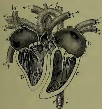
When the heart is at work, the simultaneous contraction of the two auricles (i.e. of the muscle fibres on their walls) is followed by a similar contraction of the ventricles, and this by a pause or passive interval of re-expansion, after which the rhythm of contraction recommences. In the contraction of the auricles, the mass of blood in the large veins will not permit of a backflow in a peripheral direction, so that virtually all the contents of the auricles pass into the respective ventricles, which at that moment are flaccid and uncontracted. As the ventricles fill, the valves between them and the auricles are partly closed, and this is perfectly accomplished when the contraction of the ventricle passive relaxation and expansion the diastole, and it is evident that by the systole of the auricles the ventricles are filled, and that by the systole of the ventricles their contents are for the most part forced into the arterial systems of lungs and body. As the heart usually beats about seventy-two times a minute, the cycle of events just noted lasts about ths of a second, of which the systole of the ventricle lasts about ths, that of the auricle perhaps th, and the passive interval about ths of a second.
The activity of the heart has several external indices, such as the beating, seen and felt between the fifth and sixth rib on the left side, due to contraction of the ventricles, which makes the 'apex' of the heart strike against the pericardium, and through this on the wall of the chest. There are also sounds produced by the heart: (a) the longish dull sound probably caused by the contraction of the muscular fibres of the ventricles and the tension of the valves between these chambers and the auricle; (b) the sharp sound due to the sudden closure of the semilunar valves when the contraction of the ventricles ceases. The heart sounds are of great importance in the diagnosis of disease of the heart. They may undergo various changes and may in some cases disappear or be replaced by or accompanied by murmurs. These murmurs are caused by the blood flowing through the orifices of the heart which have become changed by disease. At a distance from the heart, the pulse or regular dilatation of an elastic artery is a familiar index. The heart is under the control of three sets of nerves: (a) from ganglia in its own substance, apparently essential to the regular rhythm of contraction; (b) from the sympathetic system, apparently affecting rapidity of action; (c) from the pneumogastric or vagus nerve, coming directly from the brain, apparently with arresting power. See AORTA, ARTERY, BLOOD, CIRCULATION; textbooks of Anatomy by Quain, Turner, Mivart, Macalister, &c.; of Physiology by Foster, Huxley, Landois and Stirling, &c.
DISEASES OF THE HEART are either those affecting the various tissues composing the heart, or the nervous arrangements governing the heart.
(1) Diseases of Structures composing Heart may be primary or secondary. (a) Primary Diseases.—All the various tissues of the heart may be primarily affected. The Pericardium (sac surrounding heart) may be affected with inflammation (pericarditis). This is by no means an uncommon condition in rheumatic fever, while it also occurs in connection with some of the acute exanthemata. Fluid tends to be effused into the sac, and this produces great impairment of the heart's action. The condition frequently leads to a fatal termination. Various tumours may occur in connection with the pericardium.
The Myocardium (muscular wall of heart) may also be affected with inflammation leading to very irregular and impaired action of the heart, and often to death. This is known as myocarditis, and if the fatal termination does not ensue in the acute stage of the disease, the wall of the heart is apt to be left in a weakened condition due to the pathological changes set up in the course of the inflammation. The myocardium may occasionally be the seat of tumours. The muscular substance of the heart may undergo a fatty degeneration, which may produce death either from failure of the heart, or more rarely by rupture of the wall.
The Endocardium (lining membrane of heart; also forus the valves of the heart) is the most common seat of inflammation in rheumatic subjects and in individuals suffering from scarlet fever or from some other of the exanthemata. This endocarditis is specially apt to attack the valves of the left side of the heart, and to lead to deformity and the imperfect action of these important structures. When this occurs the well-known train of symptoms commonly associated with heart disease are apt to appear—breathlessness, palpitations, irregular heart's action, dropsy, albuminuria, &c.: while the various signs of valvular disease, among the most important of which is the alteration in the sounds of the heart, and the development of murmurs may also be determined. In many cases, however, in spite of disease of the valves, the heart may continue to act satisfactorily. But there is always a great danger of its proving inadequate to the additional work thus put upon it, and of its suddenly failing under any extra strain.
There is one peculiar form of inflammation of the endocardium known as acute ulcerative endocarditis which is exceedingly fatal, and which is due to the development of micro-organisms in the heart. Certain slow degenerative changes may also affect the endocardium, more especially where it composes the aortic valves (Atheroma).
In all inflammatory affections of the heart there is a tendency for all the structures to be involved at one and the same time. (b) Secondary Diseases.—As the result of various morbid states of other parts of the body, the heart, and more especially its muscular wall, may become secondarily affected. Thus in fever the muscular substance of the heart manifests the condition of cloudy swelling, and thus becoming weakened tends to yield to the pressure of blood inside the heart, and to undergo dilatation. This state of the organ is frequently accompanied by the develop- ment of murmurs due to the imperfect action of the valves between the anicles and ventricles allowing the blood to flow backwards through the orifices. At the same time the various symptoms of disturbed circulation are developed.
In the various forms of anæmia (bloodlessness), whether primary or secondary to other diseases, the muscle of the heart becomes debilitated, and a similar series of signs and symptoms to those just described make their appearance.
In certain diseases in which the blood pressure is raised (Bright's Disease), or when any condition throws extra work on the heart for a considerable period, the organ becomes hypertrophied—i.e. increased in size and strength. This is well seen when the valves are diseased, and the muscular substance is well nourished.
(2) Derangements of Nervous Mechanism of Heart.—As a result of many totally different conditions the sensory nervous mechanism of the heart may be affected, and give rise to pain or to various sensations in the region of the heart. These sensations are not, however, always indicative of organic disease of the organ.
A peculiar set of symptoms, known as angina pectoris, are treated more fully under a separate head. The patient suffers from attacks, the chief symptom of which is a dreadful feeling of impending death, usually with cardiac pain. When occurring as the result of organic heart disease these symptoms are most commonly connected with disease of the aortic valves.
The various nervous arrangements presiding over the movements of the heart may also become deranged, and lead to increased or diminished heart's action or to irregular action. The last is the most frequent, and is a very common accompaniment of organic disease, though it frequently occurs in individuals entirely free of any such condition. Nervous and gouty individuals and those addicted to the excessive use of tobacco are common sufferers from such palpitations.
The words 'broken heart' seem to suggest a form of heart disease. But of course the expression arose out of the long prevalent and now wholly obsolete view that the heart is in some way the seat of the affections—a view inevitably suggested by the quickening of the pulse under emotion, or its temporary stoppage from a sudden shock.