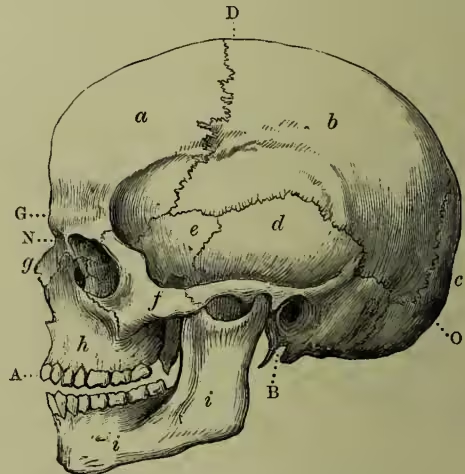
Skull. The skull is divided into two parts, the cranium and the face. In human anatomy it is customary to describe the former as consisting of eight and the latter of fourteen bones; the eight cranial bones, which constitute the brain-case, being the occipital, two parietal, frontal, two temporal, sphenoid, and ethmoid; while the fourteen facial bones, which surround the cavities of the mouth and nose and complete the orbits or cavities for the eyes, are the two nasal, two superior maxillary, two lachrymal, two malar, two palate, two inferior turbinated, vomer, and inferior maxillary. The bones of the ear, the teeth, and the Wormian bones are not included in this enumeration. The lower jaw articulates with the temporal bones by means of a diarthrodial Joint (q.v.), but all the others are joined by sutures. On the base of the cranium the occipital and sphenoid bones articulate by means of a plate of cartilage (synchondrosis) in young subjects; in adults this becomes bony union. Sutures are named from the bones between which they are found, but to those around the parietal bones special names are given—e.g. interparietal or sagittal; occipito-parietal or lambdoid; fronto-parietal or coronal; parieto-temporal or squamous. During adult life many of the sutures close by bony union and disappear, but both the age at which this occurs and the order of its occurrence are subject to variation. Wormian bones are irregular ossifications found in relation to the sutures of cranial bones, but seldom seen in relation to the bones of the face. They are most frequent in relation to the lambdoid suture, and seldom one inch in diameter. The closure of a suture stops the growth of the skull along that line, and in order to compensate for this defect an increase of growth may occur at right angles to the closed suture and thus irregularities of form may result: for example, closure of the sagittal suture stops transverse growth, but the skull continues to grow in the longitudinal and vertical directions, with the result that a boat-shaped cranium is produced—scaphocephaly. Irregular forms may be produced artificially by pressure applied early in life. This is best seen among certain American tribes who compress their children's heads by means of boards and bandages. The bones of the skull are pierced by holes (foramina), and similar holes are found in relation to the adjacent margins of bones. Most of these foramina are situated in the base or floor of the skull, and are for the ingress of arteries and the exit of veins and cranial nerves. The largest of these foramina—the foramen magnum—is found in the occipital bone. It is situated immediately above the ring of the atlas vertebra, and through it the continuity between the brain and spinal cord is established, and further, it transmits the vertebral arteries which supply blood to the brain. Compared with the skulls of animals, the form of the human skull is modified (1) by the proportionately large size of the brain and the consequent expansion of the bones which surround it; (2) by the smaller size of the face, especially of the jaws, so that the face of man, instead of projecting in front of, is under the forepart of the cranium; (3) by the crest attitude, which places the base of the skull at a considerable angle with the vertebral column, and, in consequence of a development backwards from its point of articulation with the vertebrae, the skull is nearly balanced on the summit of the vertebral column. Hence the orbits look forwards and the nostrils look downwards. The development of the skull is a subject of great interest, not only in itself, but as throwing light on many points which the study of the adult skull would fail to explain. At a very early period of fetal existence the cerebrum is enclosed in a membranous capsule external to the dura mater, and in close contact with it. This is the first rudiment of the skull, the cerebral portion of which is consequently formed before there is any indication of a facial part. Soon, however, four or five processes jut from it on either side of the mesial line, which grow downwards, incline towards each other, and unite to form a series of inverted arches, from which the face is ultimately developed. Imperfect development or ossification of these rudimentary parts of the face gives rise to 'hare-lip' and 'cleft-palate,' or in very extreme cases to the monstrosity termed 'Cyclopean,' in which, from absence of the frontal processes, the two orbits form a single cavity, and the eyes are more or less blended in the mesial line.
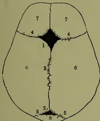
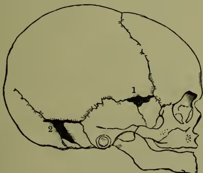
The cartilage, formed at the base of the membranous capsule, is speedily followed by the deposition of ossific matter at various points of the capsule, which soon becomes converted into flakes of bone; while the intervening portions, which remain membranous, permit the skull to expand as its contents enlarge. Then follows the appearance of ossaceous nuclei in the cartilage at the base, corresponding to the future occipital and sphenoid bones. Lastly, the various bones, some originating in membrane, and some in cartilage (see OSSIFICATION), approach one another by gradual enlargement, and become united in various ways, so as to form a continuous, and ultimately an unyielding bony case, which is admirably adapted for the defence of the brain, for sheltering the organs of special sense, and for being attached to the ligaments and muscles by which the skull is supported and moved on the spine. At the period of birth most of the principal bones have grown into apposition with their neighbours, forming the sutures; but one large vacuity remains at the meeting-point of the parietal and frontal bones, which is termed the anterior fontanelle (so called from the pulsations of the brain, which may be here seen resembling the rising of water at a spring or fountain. There are two fontanelles in the mesial line (as 1, anterior fontanelle; 2, posterior fontanelle; 3, sagittal suture; 4, 4, coronal suture; 5, lambdoid suture; 6, 6, parietal bones; 7, 7, two halves of the frontal bone, still ununited; 8, occipital bone. shown in fig. 2), and two lateral fontanelles on either side (as shown in fig. 3), which do not close till the second year after birth, and sometimes remains open much longer. The deficiency of the osseous brain-case at this position not only facilitates delivery, but also acts to some extent like a safety-valve during the first months of infantile life, at which time the brain bears an unusually large proportion to the rest of the body, and is liable to sudden variations of size from temporary congestion, and other causes. The sutures remain distinct long after the closure of the fontanelles, and serve a purpose both in permitting an increase of the size of the cranium by the growth of the bones at their edges, and in diminishing and dispersing vibrations from blows, and thus contributing to the security of the brain.
The number of centres of ossification in the skull is tolerably constant; each bone having a certain number. After the sutures have been formed, and the skull has acquired a certain thickness, a process of resorption commences in the interior of the bones, and reduces the originally dense structure to a more or less cellular or cancellated state. The interior thus altered is called the Diploë, and by this change the weight of the skull is much diminished while its strength is scarcely affected.
The diploë usually begins to be apparent about the tenth year, and is most developed in those skulls which are thickest. A continuation of the same process of resorption which causes the diploë gives rise to the formation of the cavities known as the frontal and sphenoid sinuses. The formation of the diploë divides the walls of the cranium into three layers—viz. an outer tough layer; an inner dense, brittle, and somewhat glass-like layer, known as the vitreous table or layer; and the intervening cancellous diploë. Diploë is absent from the cribriform plate of the ethmoid bone and from the roof of the orbital cavities, and thus these are the thinnest parts of the cranium. The growth of the skull after the seventh year proceeds slowly, but a slight increase goes on to about the age of twenty. The skull-bones are freely supplied with blood from arteries which pass from the dura mater internally and the pericranium externally, through the numerous foramina observed on both surfaces; the blood being returned by veins which take various directions.
The fact that concussion of the brain scarcely ever proves fatal, unless there is also fracture of the skull, affords the most distinct evidence that the skull is constructed in such a manner that so long as it maintains its integrity it is able to protect its contents from serious lesion. This marvellous protective power is due to its rounded shape, whereby its strength is increased, and in consequence of which blows tend to glide off it without doing material damage. Moreover, the curved lines or ridges which may be traced round the skull tend to strengthen it. The weakest part of the skull is at the base. Hence, notwithstanding its removal from exposure to direct injury and the protection afforded by the soft parts, fracture takes place more frequently at the base than at any other part of the skull, fracture often taking place here even when the skull was not broken at the part struck. There are two points in the architecture of the bones of the face which deserve especial notice—viz. (1) the great strength of the nasal arch; and (2) the immobility of the upper jaw, which is fixed by three buttresses—the nasal, the zygomatic, and the pterygoid.
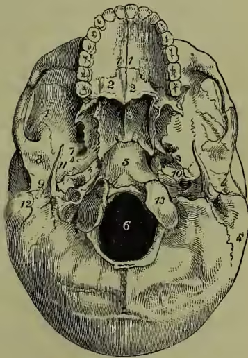
The base of the skull, whether seen from within or from below, presents many objects of physiological interest in relation to the nervous system. As seen from within, the base presents on each side three fossæ, corresponding to the anterior and middle lobes of the cerebrum and to the cerebellum. These 1, 1, hard palate; 2, 2, palate bones; 3, vomer; 4, zygomatic fossa; 5, basilar process of the occipital bone; 6, foramen magnum; 7, foramen ovale; 8, glenoid fossa; 9, external auditory foramen; 10, carotid foramen of the left side; 11, styloid process; 12, mastoid process; 13, one of the condyles of the occipital bone. fossæ are marked, as is the whole skull-cap, by the cerebral convolutions, and they contain numerous 'foramina' and 'fissures' which give passage to various sets of nerves and blood-vessels. The external or outer surface of the base of the skull, if we consider it from before backwards, is formed by the palate processes of the superior maxillary and palate bones; the vomer; the pterygoid and spinous processes of the sphenoid and part of its body; the under surface of the temporal bones; and the occipital bone. The most important of the parts which it presents are named at fig. 4.
The Morphology of the Skull is the highest and most difficult problem of comparative anatomy, and has cost the most extraordinary labour for its solution. Goethe and Oken independently suggested that the skull was to be regarded as the modification of a series of four vertæbræ, and this 'vertebral theory' was worked out in the most elaborate detail by Owen and other anatomists (see SKELETON). Huxley, however, in a celebrated Croonian Lecture (1858), revised and extended the hitherto neglected embryological observations of Rathke, proposed an unanswerable destructive criticism of the archetypal theory, and may be said to have thus definitely placed the newer view in the way of general acceptance. An enormous amount of detailed research, for which we are indebted chiefly to Parker in England and Gegenbaur in Germany, has established the newer theory on the sure ground of actual observation.
Taking first the simple unsegmented cartilaginous cranium of a skate or dog-fish, with its appended jaws and branchial arches, we find that in development, though the notochord extends into the region of the head, the vertæbræ stop altogether short of it; but that on each side of the cranium there arise a pair of cartilaginous bars—the trabeculæ or 'rafters' of the future skull—and three pairs of cartilaginous capsules, nasal, ocular, and auditory, form round the developing sense-organs; the nasal capsules immediately unite with the ends of the trabeculæ, which are meanwhile uniting below, and growing up at the sides to form the brain-case. The auditory capsules become united with the trabeculæ by the appearance of two new masses of cartilage—the parachordals—the eyes of course remaining free. At first there are no jaws, but a series of seven or more similar vertical cartilaginous bars or arches, considerably resembling the trabeculæ, between which slits open into the pharyngeal cavity. The first pair of these arches develops an ascending process, which passes above the developing mouth, and becomes the 'palate-pterygoid' arch or upper jaw, the original portion remaining as the mandible. The second pair of arches—the 'hyoid'—becomes more or less modified usually to aid in supporting the jaws and floor of the mouth, while the remaining pairs become little modified, and serve throughout life to support the gills.
The more complex bony skulls of higher vertebrates are now in principle readily understood. The chondro-cranium and subjacent arches in all cases develop in the same way, although reduction and even atrophy of the gill arches subsequently takes place. The bones, although similar in the adult, originate in two utterly distinct ways, either by actual ossifications in the substance of the chondro-cranium and jaws, or by the ossification of overlying dermis, and are hence known as cartilage bones and membrane bones respectively—the latter corresponding to the dermal bones and teeth of ganoid and elasmobranch fishes. In mammals a further extraordinary specialisation takes place: the ends of the mandibular and hyoid arches lose their suspensory function, are taken up during development into the interior of the ear capsule, and are metamorphosed into the auditory ossicles.
Various Forms of the Skull.—Age.—At birth the existence of the fontanelles has already been referred to. The frontal and parietal eminences are especially prominent, and the mastoid process is absent. The face is only one-eighth of the bulk of the cranium, whereas in the adult the face is equal to one-half. During the first seven years the skull grows rapidly, and by this time many parts have attained definite size. At the period of puberty the face and regions of the air-sinuses undergo expansion. The face elongates owing to the growth of teeth and the increase in the size of their alveolar sockets. In old age the skull may become lighter and thinner or the reverse. Loss of the teeth and absorption of their sockets result in diminution of the size of the face, and thus the upper jaw recedes, while the chin becomes prominent.
Sex.—It is not always possible to determine the sex from the skull; but, as a rule, the skull of the male has more strongly marked muscular impressions, while the mastoid processes, superciliary ridges, and air-sinuses are more pronounced than in the female, whose skull generally retains the leading features of a young skull.
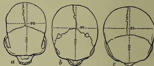
Race.—In comparing the skulls of different races of mankind it is necessary to have recourse to various methods of measurement, and these are usually conducted on the skulls of adult males. The following is a short summary of these methods. (a) Cranial Capacity.—This is obtained by filling the cranial cavity with shot, and then measuring the quantity in a graduated vessel, special precautions being observed in order to obtain equable results. The capacity of normal human crania varies from 60 to 110 cubic inches—the average in all races being 85 cubic inches—e.g. Eskimo, 91.5; European, 90.3; Chinese and Mongols, 87.3; African Negroes, 82.4; Native Australians (aboriginal), 79.3; Andaman Islanders, 78.1. (b) Linear measurement of the horizontal circumference of the cranium. In the adult European male the average is 20.7 inches, and in the female 19.6 inches. (c) A third method is by comparison of the relative length, breadth, and height of the cranium. The standard of maximum length is taken as 100, and thus ; and on this basis skulls are classified in three groups—viz.:
Brachycephalic = breadth-index above 80.
Mesaticephalic = " " from 75 to 80.
Dolichocephalic = " " below 75.
In a similar way the proportion of height to length may be calculated, and a height-index established. It varies less than the breadth-index—e.g.:
| Breadth. | Height. | |
|---|---|---|
| Mongolians of Siberia and Central Asia..... | 88 | 73 |
| Andaman Islanders ..... | 82 | 77 |
| Chinese ..... | 79 | 75 |
| English ..... | 76 | 71 |
| Native Australians (aboriginal)..... | 71 | 71 |
| Fiji Islanders..... | 66 | 74 |
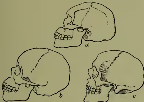
a, Australian, prognathous; b, African, mesognathous;
c, European, orthognathous. (After Tylor.)
(d) The Degree of Projection of the Jaws.—We have seen that the human skull, when compared with the skulls of lower animals, presents a small face extended vertically, and thus placed under the anterior part of the cranial box. Prominent jaws therefore indicate an approach to an animal type, especially when associated with a receding forehead. The degree of projection is expressed by the gnathic index of Flower, and is obtained by comparing the basi-alveolar length with the basi-nasal length. When the gnathic index is below 98, skulls are said to be orthognathous; from 98 to 103, mesognathous; above 103, prognathous—e.g.:
| Gnathic Index. | |
|---|---|
| English ..... | 96 |
| Chinese..... | 99 |
| Eskimo..... | 101 |
| Fiji Islanders ..... | 103 |
| Native Australians (aboriginal) ..... | 104 |
(e) The form of the nasal skeleton and its anterior openings is also subject to variation, and so its height and width may be measured, and the relation between the two expressed as a nasal index.
Nasal index below 48 = leptorhine.
" from 48 to 53 = mesorhine.
" above 53 = platyrrhine.
For example:
| Nasal Index. | |
|---|---|
| Eskimo..... | 44 |
| English..... | 46 |
| Chinese..... | 50 |
| Native Australians (aboriginal)..... | 57 |
In a similar way the form of the orbit is subject to variation, but this is of less consequence than the nose. Other measurements of the face skeleton are also made. A time-honoured measurement, long thought to be sufficient in itself for founding a classification of races, was the Facial Angle of the Dutch anatomist Peter Camper (q.v., 1722-89). This was obtained by drawing one line from the centre of the forehead to the most projecting part of the upper jaw just above the incisor teeth, and another from the opening of the ear to the base of the nasal opening; between these was contained the facial angle.
For the relation of craniometry to the science of man, see ETHNOLOGY and works there cited. See also the articles ANTHROPOMETRY, ANTHROPOID APES, BRAIN, MAMMALS, MAN, PHRENOLOGY, SKELETON, VERTEBRATA; Huxley's Croonian Lecture (1858); Huxley's Anat. of Verteb. Animals (1871); Parker's Morphology of the Skull (1877); and for summary, Balfour's Embryology (vol. ii.); Broca, Instructions Craniologiques et Craniométriques; Flower, Cat. of Mus. of Roy. Coll. Surg. of Eng. (part i. 1873-79); Turner, Challenger Reports, Zoology, x. (1888); Schmidt, Anthropologische Methoden (1888); Benedikt, Kraniometrie und Kephalometrie (1888). For the illustrative tables in this article we are indebted to the summary in Quain's Anatomy (10th ed.).