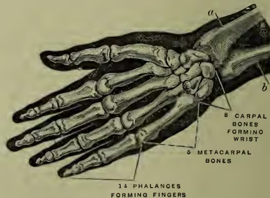
a, radius; b, ulna.
Hand, THE. The genus Homo, or Man, was ranked by Cuvier in his classification of mammals as a distinct order, Bimana, in consequence of man being the only animal possessing two hands. Recently the tendency has been to revert to the classification of Linnaeus, and to place man with all monkeys, lemurs, and bats in the order Primates (see BIMANA, MAMMALIA). At first sight it might be considered that the so-called Quadrimana or four-handed animals (monkeys, &c.) were better equipped than those which possess only two hands, but this is far from being the case. None of the four hands are adapted to the variety of actions which the human hand is capable of performing, and they are all, to some degree, required for support and locomotion; so that, while in the higher forms of the quadrimana the extremities present an approximation in structure to those of man, in the lower they gradually tend to resemble the ordinary quadrupedal type. 'That,' says Cuvier, 'which constitutes the hand, properly so called, is the faculty of opposing the thumb to the other fingers, so as to seize upon the most minute objects—a faculty which is carried to its highest degree of perfection in man, in whom the whole anterior extremity is free, and can be employed in prehension.' The peculiar prehensile power of the human hand is chiefly dependent upon the length, power, and mobility of the thumb, which can be brought into exact opposition to the extremities of all the fingers, whether separately or grouped together.
The general arrangement of the bones of the hand will be understood by a reference to fig. 1.
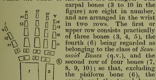
In fig. 2 we have a diagram showing the way in which the bones of the hand are arranged. The 1, end of radius; 2, end of ulna; 3, scaphoid; 4, semilunar; 5, cuneiform; 6, pisiform; 7, trapezium; 8, trapezoid; 9, magnus; 10, unciform; 11, metacarpal bones; 12, 13, 14, first row of phalanges; 1, thumb; II, forefinger, &c.; V, little finger. carpal bones (3 to 10 in the figure) are eight in number, and are arranged in the wrist in two rows. The first or upper row consists practically of three bones (3, 4, 5), the fourth (6) being regarded as belonging to the class of Sesamoid Bones (q.v.), and the second row of four bones (7, 8, 9, 10); so that, excluding the pisiform bone (6), the carpal and the tarsal bones correspond in number. As we commonly term the palm the front of the hand, the thumb becomes conventionally the outer, and the little finger the inner digit; but according to the rules of comparative anatomy, and in order to compare the hand and foot, we ought to reverse these terms. The outer (3) of the carpal bones of the first row supports (through the intervention of 7 and 8) the bones of the thumb and forefinger (I and II), and constitutes with them the outer division of the hand. The inner (5) of the carpal bones bears the little and the next (the ring) finger (V and IV), and constitutes with them the inner division of the hand, while the middle one (4) bears the middle finger (III), and belongs to the middle division of the hand. We likewise see from this figure, and also from fig. 1, that the two outer bones (3 and 4) are connected with the radius, while the inner bone (5) is connected (indirectly by a thick ligament) with the ulna.
The carpal bones are so arranged that the carpus presents a dorsal convex surface, upon which the tendons of the extensor muscles of the fingers play, and a palmar concave surface on which the tendons of the flexor muscles lie. The several bones are joined to one another—each bone being united to three or more others—by a large extent of surface, and are girded together by strong ligamentous bands. The wrist is thus as strong as if it had been constructed of one solid piece of bone, while the slight gliding movements which occur between the several bones give it an elasticity which serves to break the shocks that result from falls upon the hand. The uppermost surface of the first row of carpal bones is convex, and this convex surface is received into a wide cup or socket, formed by the lower articular surface of the radius and by a ligament passing from that bone to the ulna. Like the great toe, the thumb has only two phalanges, while each of the other digits has three.
For the different directions in which the arm and hand collectively can be moved, see the description of the construction and movements of the shoulder and elbow joints at ARM. Movements of the forearm and hand, to which there is virtually nothing analogous in the leg, are those of 'pronation and supination.' In pronation (derived from pronus, 'with the face downwards') we turn the palm of the hand downwards, as in picking up any object from the table; in supination (derived from supinus, 'with the face upwards'), we turn the palm upwards, as for the purpose of receiving anything that may be placed in it.
These movements of pronation and supination are so important to the usefulness of the hand that we must notice the muscles by which they are chiefly effected. One of these muscles passes from a projecting process on the inner side of the arm-bone at its lower end to the outer edge of the middle of the radius. Its contraction causes the radius to roll over, or in front of, the ulna. It thus pronates the hand, and is called a pronator muscle. Another crosses from the front of the lower end of the ulna to the corresponding part of the radius. Its shape and its action are indicated by the name pronator quadratus. Another muscle passes from a projecting process on the outer side of the arm-bone and from the outer aspect of the ulna to the outer surface of the radius near its upper part. It runs therefore in an opposite direction to the former muscle, and produces an opposite effect, rolling the radius and the hand back into the position of supination. Hence it is called a supinator muscle (see fig. 3). The fourth is a very powerful muscle termed the Biceps (q.v.), which not only bends the elbow, but, from the mode in which its tendon is inserted into the inner side of the radius, 'also rotates the hand; and it gives great power to that movement. When we turn a screw, or drive a gimlet, or draw a cork, we always employ the supinating movement of the hand for the purpose; and all screws, gimlets, and implements of the like kind are made to turn in a manner suited to that movement of the right hand, because mechanics have observed that we have more power to supinate the hand than to pronate it.' Supination can only be performed to its full extent by man, and even in man it is not the natural or habitual position; monkeys can partially effect the movement, and in most of the lower animals the part corresponding anatomically to the hand is constantly in a state of pronation.
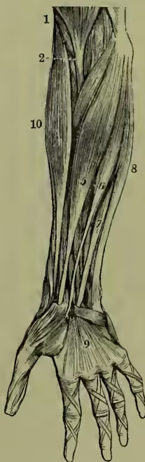
The movements of which the hand itself, without reference to the arm, are capable, are very numerous, and in this respect differ considerably from the corresponding movements of the foot. Thus we can bend the fingers down upon the palm, or we can extend them beyond the straight line; we can separate them from one another to a considerable extent, and we can close them with considerable force. The wrist and hand are bent forwards or flexed upon the forearm by three muscles which pass downwards from the inner 1, biceps; 2, tendon of biceps; 5, the radial flexor of the wrist; 6, the long palmar muscle, spreading out (at 9) into the palmar fascia; 8, the ulnar flexor of the wrist; 10, the long supinator muscle. condyle or expanded end of the humerus, and are termed the radial flexor, the ulnar flexor, and the long palmar muscles. The first two of these muscles are inserted into wrist-bones on the radial and ulnar sides respectively, while the third expands into a fan-like fascia or membrane in the palm of the hand, and thus serves both to support the skin of the palm and to protect the nerves and vessels which lie below it. Beneath the palmar fascia lie two sets of flexor muscles of the fingers, and they present so beautiful a mechanical arrangement as to merit special notice.
The superficial or perforated flexor muscle passes down the front of the forearm, and divides into four tendons, which become apparent after the removal of the palmar fascia, and are inserted into the second phalanges of the fingers, each tendon splitting at its termination, to give passage to the similar tendons of the deep or perforating flexor muscle, which passes from the upper part of the ulna to be inserted into the last phalanx of each finger. This arrangement of the tendons of the superficial and deep flexor muscles is shown in fig. 4. To these flexor muscles
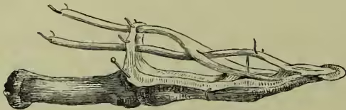
To show the perforation of one of the tendons of the superficial flexor muscle (which is inserted into the second phalanx), in order to allow the corresponding tendon of the deep flexor to pass onwards to be inserted in the last phalanx. correspond the common extensor muscle of the fingers, which, like the flexors, divides into four tendons, one for each finger. Besides these, there is a special extensor of the index-finger, a series of muscles forming the ball of the thumb, which move that organ in almost every direction, and various small muscles giving lateral and other movements to the fingers.
It is sufficient to observe that the hand is very richly supplied with blood-vessels and nerves, without entering into any anatomical details on these points. There is no part of the body where the sense of touch is so acute as at the tips of the fingers; but we defer to the article TOUCH the consideration of the special arrangements which make this part of the hand peculiarly important in relation to our knowledge of external objects.
As a measuring standard for the height of horses a hand is a palm-breadth, assumed to be four inches. For left-handedness, &c., see RIGHT- AND LEFT-HANDEDNESS.
Our notice of the comparative anatomy of the Foot (q.v.) renders it unnecessary to trace the modifications presented in the lower animals by the bones corresponding to those of the human hand, as the carpal and metacarpal bones with their phalanges undergo adaptations of form to meet the individual wants of the animal, very much in the same manner as the tarsal and metatarsal bones and their phalanges. Thus, the reader will readily see that the so-called knee of the horse, for example, is the carpus, and he will have no difficulty in tracing the metacarpal bones and phalanges. See Sir Charles Bell, The Hand, its Mechanism and Vital Endowments (Bridgewater Treatise, 1836; 9th ed. 1874).